Chapter 18 Hysteroscopic Myomectomy and Polypectomy, and Removal of Retained Products of Conception. Operative Techniques
Chapter 18 Hysteroscopic Myomectomy and Polypectomy, and Removal of Retained Products of Conception
GENERAL PRINCIPLES
Definition
■ Uterine leiomyomas also called uterine fibroids, myomas, or fibromyomas are benign proliferative, well-circumscribed, pseudoencapsulated benign growths composed of smooth muscle and fibrous connective tissue. They are the most common benign growth of the uterus. These benign growths may be located in the body of the uterus and cervix including endocervical, intracavitary, submucosal, intramural, transmural, subserosal exophytic may pedunculated positions, and may prolapse through the cervix. The size, number, and location of fibroids are unique to each patient and may be associated with a variety of clinical symptoms or menstrual aberrations. The pathogenesis of leiomyomas remains unknown.
■ Endometrial polyps are benign growths of the endometrium. They are common throughout the lifespan of women. Most endometrial polyps are asymptomatic. Generally, they are single and may occur anywhere within the uterine cavity or near the tubal ostia. Polyps may occur also within the endocervix and ectocervix. They are usually single, sessile, but may be on a stalk, pedunculated, or prolapse through the ectocervix. In reproductiveaged women, the risk of coexisting endometrial hyperplasia (simple, complex without atypia, or complex hyperplasia with atypia) or malignancy within a polyp is approximately 1.7%. However, among women older than 60 years of age with symptomatic postmenopausal bleeding, there is an 8.3- fold increased risk of premalignant changes. Symptomatic polyps detected during the menopause may be associated with a 5% risk of malignancy. The pathogenesis of endometrial polyps remains elusive; however, increased risk is noted in women who use tamoxifen, are overweight/obese, hypertensive, diabetic, and associated with hormone replacement therapy.
■ Retained products of conception can be seen following miscarriage, termination of pregnancy (first or second trimester), anembryonic firsttrimester miscarriage, incomplete miscarriage, missed abortion, postpartum hemorrhage with “blind suction D&C,” vaginal delivery, manual removal ofplacenta, C/section, or in pregnancies complicated by Müllerian anomalies.
■ Most often patients are counseled to undergo expectant management which is associated with 81% success.
■ However, one out of five women may have persistent bleeding, cramping, leukorrhea, fever, abdominal pain, incessant menstrual bleeding, or transvaginal sonographic findings suggestive of RPOC (homogeneous or heterogeneous echogenic foci or endometrial fluid collection with echogenic foci, coupled with high-velocity, low-resistance flow at color Doppler ultrasonography).
■ Among women who become pregnant after vigorous curettage, there is an increased risk of abnormal placentation in future pregnancies predisposing patients to placenta accreta/increta/percreta.
■ For these women, historically, a “blind D&C” with vacuum aspiration has been performed. However, surrounding healthy, normal, viable endometrial tissue may be altered leading to Asherman’s syndrome (mild, moderate, or severe) or resultant hypomenorrhea due to extensive alteration of the endometrial basalis layer.
Differential Diagnosis
■ Endometrial polyps
■ Adenomyomatous polyp
■ Leiomyosarcoma
■ Endometrial stromal tumor
■ Stromal tumor of uncertain malignant potential
■ Calcified retained products of conception
■ Intracavitary endometrial blood clots
■ Intracavitary leiomyoma
■ Atypical leiomyoma
■ Adenomyoma
Anatomic Considerations
■ The FIGO classification system is useful in determining the position of the uterine fibroid within the endometrium and the depth of penetration into the myometrium.
■ Hysteroscopic removal of uterine fibroids in general is limited to patients with FIGO classification type 0 and type 1 leiomyomas. Expert hysteroscopic surgical experience may permit removal of small type 2 leiomyomas (Fig. 18.1).
■ Type 0 leiomyomas are entirely within the uterine cavity with no myometrial extension. The base can be pedunculated, narrow, or wide(Fig. 18.2).
■ Type 1 leiomyomas involve <50% of the myometrium. When viewed hysteroscopically, there is >90-degree angle of the leiomyoma surface to the uterine wall (Fig. 18.3).
■ Type 2 leiomyomas involve >50% myometrial extension. When viewed hysteroscopically, there is a <90-degree angle of the leiomyoma surface to the uterine wall (Fig. 18.4).
■ While some type 2 leiomyomas may be performed hysteroscopically, expert surgical experience is needed. More commonly, they are removed by a laparoscopic/robotic or laparotomic approach.
■ It is important to determine the distance from the outer edge of the leiomyoma to the serosa with ultrasound or pelvic MRI with and without contrast to determine if a hysteroscopic approach is feasible.

Figure 18.1. FIGO classification of types 0 and 1 fibroids.
■ The myometrium remodels during and after hysteroscopic myomectomy. It is advisable to exclude hysteroscopic resection of a leiomyoma that is within 1.0 cm from the serosal edge. Adherence to this guideline decreases the risk of uterine perforation.

Figure 18.2. MRI with well-defined FIGO type 0 fibroid.

Figure 18.3. SIS demonstrates FIGO type 1 fibroid.
■ Fluid absorption during hysteroscopic myomectomy is influenced by length of surgery, intrauterine pressure, fibroid size, number of fibroids treated, depth of myometrial involvement, breach of myometrial venous sinuses during surgery, and less significantly transtubal reflux.
■ The myometrium contains many venous sinuses. When these are opened by resection or morcellation technique, the fluid used during hysteroscopy is absorbed intravascularly. Thus, increased risk of fluid absorption occurs when type 1 and type 2 leiomyomas are treated.
■ Exceeding fluid absorption guidelines is associated with risks unique to the type of fluid used (isotonic or nonisotonic fluid).

Figure 18.4. MRI depicts FIGO type 2 fibroid.
■ Leiomyoma size, depth of myometrial involvement, number and location of intracavitary fibroids, and surgical expertise determines feasibility, safety, and ability to perform hysteroscopic removal as a single surgical procedure.
■ Some hysteroscopic myomectomy procedures will require a two-stage procedure due to the inability to complete the initial surgery due to excessive fluid absorption.
■ With expert preoperative evaluation, the informed consent will reflect the discussion regarding the possibility of incomplete or two-staged hysteroscopic treatment.
■ As the size of the leiomyoma increases, so does the volume of resected tissue. This affects the length of surgery, amount of fluid used, ability to complete the hysteroscopic resection, and risk of surgical complications.
■ The volumetric formula that describes the volume of hysteroscopic tissue removed is:
■ 4/dr3
■ 1 cm = 1/2 cm3 tissue
■ 2 cm = 4 cm3 tissue
■ 3 cm = 14 cm3 tissue
■ 4 cm = 33 cm3 tissue
■ Anatomic and surgical considerations particular to hysteroscopic resection include:
■ Presence of blood, clots, and endometrial tissue debris
■ Endometrium (especially secretory or exaggerated proliferative endometrium)
■ Intracavitary bubbles
■ Tissue or “chip” management
■ Ability to anatomically recognize the pseudocapsule and myometrial
fascicles
■ Navigation within the uterine cavity and determining the depth of fibroid resection
■ Uterine walls collapsing and increased juxtaposition of the uterine walls as hysteroscopic resection progresses
■ Fluid absorption
■ Uterine perforation
■ Uterine distensibility
■ C/section scar
■ Uterine size
■ Uterine position
■ Retroversion
■ Retroflexed
■ Axial
■ Deviated
■ Fixed position
■ Cervix
■ Cervical perforation
■ Cervical stenosis
■ With multiple insertions and removal of the hysteroscope in women with a stenotic cervix, risk of cervical lacerations, creation of false tracks, or uterine perforation may occur.
■ Tortuous cervical canal
■ Cervical laceration
■ Creation of cervical false tracks
■ The cervix may become more patulous with multiple insertions of the hysteroscope making it more difficult to maintain intrauterine pressure and intrauterine distention.
■ Placement of additional cervical tenaculum may be required to occlude a patulous cervix.
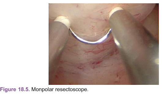
Figure 18.5. Monpolar resectoscope.
■ Hysteroscopic removal of leiomyomas can be accomplished with a hysteroscopic resectoscope: monopolar or bipolar devices (Figs. 18.5 and 18.6).
■ Increasingly, hysteroscopic morcellators are available and utilize saline as the distension medium and are more commonly employed for type 0 leiomyomas (Figs. 18.7 and 18.8).
■ Complete removal of type 0 and type 1 fundal leiomyomas and fundal endometrial polyps is more difficult with a hysteroscopic morcellator.
■ It is more difficult because the fibroid or polyp is flushed with the fundus and the morcellator operating device can not conform to reach the fundal location as well. Use of intermittent uterine decompression may facilitate complete removal.
■ Increased surgical expertise and experience permits removal of deeper leiomyomas.

Figure 18.6. Bipolar resectoscope; note the golden orange halo produced by the energy source.■ Ideally, both hysteroscopic resection surgical expertise and hysteroscopic morcellation should be within a surgeon’s armamentarium, as anatomic variations define choice of surgical equipment and fluid used.

Figure 18.7. Smith & Nephew hysteroscopic morcellator equipment.
Nonoperative Management
■ The prevalence of uterine fibroids varies between 20% and 80% in the female population. Risks of developing uterine fibroids are influenced by age, ethnicity, family history, and parity.
■ The majority of women with uterine fibroids are asymptomatic.
■ Surgery should not be offered to asymptomatic women, unless the location of the fibroid is clearly associated with infertility or impair fertility treatments such as in vitro fertilization (IVF).
■ Among patients with recurrent pregnancy loss and preterm labor consultation with a maternal fetal medicine physician or reproductive infertility physician is advisable to determine if the surgical removal would improve pregnancy outcomes.
■ Medical therapy for heavy menstrual bleeding can include a trial of tranexamic acid (for women with ovulatory heavy menstrual bleeding without thromboembolic risk factors), low-dose oral contraceptive pills, or nonsteroidal medications.

Figure 18.8. Hologic hysteroscopic morcellator equipment.
■ Intrauterine contraceptive progesterone containing devices should not be placed in women with known intracavitary fibroids as it may be associated with increased risk of IUD expulsion, inaccurate placement, malposition, or uterine perforation.
■ Patients may also have coexisting anovulatory cycles with intracavitary fibroids or endometrial polyps. Medical therapy for anovulatory may also need to be included.
■ Expectant management
■ Anatomic consideration for endometrial polyps differs from uterine fibroids because endometrial polyps are soft growths attached to the endometrium, endocervix, or ectocervix.
■ More commonly, they are single; however, they can be multiple.
■ May be associated with other intracavitary pathology including uterine fibroids, endometrial hyperplasia, or malignancy.
■ If they are asymptomatic and found coincidentally, they usually do not need to be removed.
■ Endometrial polyps associated with infertile patient should be removed hysteroscopically.
■ Patients who develop endometrial polyps while using tamoxifen and have abnormal uterine bleeding or leukorrhea should have them removed.
■ Removal of endometrial polyps should be performed with a hysteroscopic approach to increase the chance of complete removal.
■ Blind approaches for removal of endometrial polyps such as dilation and curettage are associated with incomplete resection and remnants.
■ Incomplete resection may be associated with recurrent or persistent symptoms.
■ Endometrial polyps may be removed with hysteroscopic wire loopresection or a hysteroscopic morcellator. They should not be treated with desiccation technology because tissue sampling would not be possible.
■ In general, hysteroscopic polypectomy procedures have the same risks as noted in hysteroscopic removal of uterine fibroids.
■ The same surgical principles and considerations should be followed as hysteroscopic myomectomy.
■ Endometrial polyps do not involve the myometrium; therefore, hysteroscopic resection or morcellation of polyps should be limited to the endometrium only.
■ When asymptomatic endometrial polyps are coincidentally found with TVUS, they can be followed for a short length of time 6 to 12 months with repeat TVUS.
■ If there are no clinical symptoms including pelvic pain, leukorrhea, vaginal spotting, or heavy menstrual bleeding, then imaging can be discontinued.
■ If the endometrial echo increases, hysteroscopic surgical removal is recommended.
■ Some endometrial polyps regress over time.
IMAGING AND OTHER DIAGNOSTICS
■ Evaluation of the endometrial cavity in patients with suspected intracavitary fibroids, endometrial polyps, and retained products of conception may include:
■ office hysteroscopy;
■ diagnostic hysteroscopy in an office or ambulatory surgical center;
■ transvaginal ultrasound (TVUS);
■ saline infusion sonography (SIS) with 2D transvaginal ultrasound;

Figure 18.9. Diagnostic hysteroscopy reveals fundal myoma.■ 3D saline infusion images with transvaginal ultrasound;
■ MRI of the pelvis with and without contrast (not indicated for primary evaluation of endometrial polyps and retained products of conception unless placental accreta/percreta/increta is suspected).
■ Diagnostic hysteroscopy can determine the presence of type 0, type 1, and some type 2 leiomyomas (Fig. 18.9).
■ Type 1 and type 2 leiomyomas viewed hysteroscopically can be suspected by the angle of inclination of a fibroid that abuts the endometrial cavity.
■ However, if the size of the entire fibroid and the depth of myometrial penetration cannot be ascertained by hysteroscopy, additional imaging is needed.
■ If there is uncertainty about the depth of myometrial involvement, utilize SIS with 2D or 3D ultrasound.
■ The 3D SIS coronal view is extremely useful in determining the depth of leiomyoma penetration.
■ When 2D or 3D SIS is unsatisfactory or cannot be performed, then MRI of the pelvis with and without contrast helps delineate the boundaries of the leiomyoma.
■ Endometrial polyps are well visualized during diagnostic hysteroscopy, 2D SIS, and 3D SIS ultrasound.
■ MRI with and without contrast may be considered when bimanual uterine fibroid size is greater than 12 to 14 gestational week size, limited uterine distention with SIS, or in patients who do not tolerate pelvic examinations.
■ Consider MRI when there are symptoms and signs of adenomyosis:
■ Boggy and tender uterus on clinical examination
■ Significant dysmenorrhea
■ Dysmenorrhea and irregular menstruation
■ MRI of the pelvis may be considered in virginal patients and those who do not tolerate pelvic examinations and transvaginal imaging.
■ MRI of the pelvis is useful in cases where uterine distention is limited including:
■ Patulous cervix
■ Large intracavitary lesions
■ Prior endometrial ablation
■ Adenomyosis
■ Uterine size greater than 12 to 14 weeks
■ Limited view of the endometrium due to copious bleeding which is associated with increased false positive results.

Figure 18.10. Two-dimensional saline-infused sonogram, sagittal view, demonstrates FIGO type 1 fibroid.
■ MRI is not indicated for the evaluation of endometrial polyps. While endometrial polyps may be visualized when MRI is performed for uterine fibroids, the expense of MRI and decreased sensitivity make it impractical for clinical use.
■ While TVUS is helpful in determining the presence of uterine fibroids, the location and depth of myometrial penetration are more difficult. Therefore, we recommend 2D and 3D SIS for more accurate characterization of leiomyomas (Figs. 18.10 and 18.11).
■ SIS with both 2D and 3D images helps define the topography of the leiomyoma including: size, number, location, and depth of myometrial penetration of the leiomyoma.
■ SIS has greater sensitivity and specificity compared to TVUS in detecting endometrial pathology.
■ In patients who have an inconclusive TVUS SIS is advisable.
■ Endometrial polyps are well imaged with 2D SIS and 3D SIS.
■ Accurate preop determination of the topography of intracavitary fibroids, endometrial polyps, and retained products of conception enhances surgical informed consent; predicts length of surgery, surgical expertise needed, likelihood of incomplete hysteroscopic resection/morcellation, complications, selection of hysteroscopic equipment and fluid needed (Fig. 18.12).

Figure 18.11. Three-dimensional, saline-infused sonogram with FIGO type 2 fibroid.

Figure 18.12. Intracavitary distention of fluid or CO2 may flatten lesions and make them disappear, often referred to as “the disappearing act.”
PREOPERATIVE PLANNING
■ The preoperative planning caveats and principles discussed in this sectionare inclusive to patients being treated for hysteroscopic myomectomy, hysteroscopic polypectomy, and hysteroscopic treatment of retained products of conception.
■ Operative hysteroscopy requires continuous infusion of fluid in order to distend the uterine cavity and provide clear visualization during surgery.
The amount of fluid absorption and length of surgery will vary depending on the pathology encountered.
■ Pulmonary, cardiac, and renal status should be assessed prior to surgery.
■ While blood loss is minimal during most hysteroscopic surgical procedures it is important to replete iron stores prior to surgery. This can be accomplished with oral iron supplementation or IV iron infusion symptomatic patients.
■ Hysteroscopy should not be performed if a patient has active pelvic inflammatory disease, acute endometritis, active herpes infection, or pyometra.
■ Hysteroscopy should not be performed in febrile patients whose source of fever is attributable to a tubal–ovarian abscess, pelvic inflammatory disease, or acute endometritis.
■ Patient should not have a viable intrauterine pregnancy.
■ When possible, hysteroscopy should be scheduled during the proliferative phase of the menstrual cycle due to improved visualization.
■ Operative hysteroscopic surgery should not be canceled in women that are actively bleeding.
■ The intraoperative use of a fluid management system irrigates blood, blood clots, and debris, and provides uterine distention.
■ The ability to vary the intrauterine pressure can facilitate visualization by tamponade of endometrial/myometrial arterioles in patients who are actively bleeding.
■ Intracervical injection of dilute vasopressin may decrease active bleeding.
■ Consider misoprostol prior to hysteroscopic surgery because it:
■ facilitates cervical dilation;
■ decreases risk of cervical lacerations;
■ decreases risk of creating of false tracks;
■ decreases risks of uterine perforation;
■ enhances myometrial contractility.
■ Misoprostol-associated myometrial contractions may cause a type 1 leiomyoma become a type 0 or facilitate complete resection of deeper leiomyomas as they are pushed into the endometrial cavity.
■ Occasionally a type 0 leiomyoma may fully prolapse through theectocervix and vaginal myomectomy can be performed.
■ Mix 20 units of vasopressin in 200 mL normal saline and inject in 5 mL aliquots into the intracervical stromal (1 cm depth) at 11, 2, 4, and 8 o’clock position.
■ Prior to intracervical injection, confirm with the anesthesiologist that patient is hemodynamically stable.
■ Aspirate and inject the dilute vasopressin slowly into the cervical stroma. Do not inject when blood is aspirated.
■ Monitor vital signs closely during the injection as vasopressin may cause bradycardia, cardiac arrhythmias, hypertension, and death.
■ Inform the anesthesiologist of the total amount of vasopressin used.
■ Perform a pelvic examination and visualize the cervix in the office prior to scheduling surgery. This allows the surgeon to anticipate potential difficulties intraoperatively with cervical dilation or visualization of the cervix.
■ In addition to prescribing misoprostol for cervical priming, the surgeon may also want to have a transabdominal ultrasound available to facilitate safe cervical dilation in potentially difficult cases.
■ Cervical stenosis can be anticipated under the following circumstances:
■ Difficulty in performing a pap smear
■ Apical vaginal agglutination
■ The cervix is flushed against the vaginal vault
■ Prior LEEP or cone biopsy
■ Nulliparity
■ Menopausal status with atrophic vaginal changes
■ Prior Cesarean sections
■ Consider a two day preoperative course of oral or vaginal misoprostol prior to operative hysteroscopy to facilitate cervical dilation.
■ Prescribe misoprostol 400 mcg by mouth or vaginally at bedtime 2 days before surgery and misoprostol 400 mcg by orally or vaginally at bedtime the night before surgery.
■ Side effects of misoprostol may include nausea, uterine cramping, pelvic pain vaginal bleeding, diarrhea, or pyrexia.
■ Patients may take an NSAID to mitigate these side effects.
■ Historically laminara has been used as an osmotic cervical dilator; however, it has become less practical due to the need for an additional office visit for placement. Additionally, attempts at placement of an extra thin laminara may not be possible with marked cervical stenosis.
■ A shallow or “mini” LEEP cone biopsy may be needed to excise the ectocervix in order to successfully dilate the endocervix.
■ Preoperative anticipation of a difficult dilation allows the surgeon to request ancillary equipment such as the LEEP machine and disposable LEEP wire loop.
■ Informed consent for a LEEP procedure should be obtained when cervical stenosis is anticipated.
■ Consider intraoperative transabdominal ultrasound guidance when marked cervical stenosis is anticipated. This permits real-time transabdominal ultrasound guidance during cervical dilation and decreases the creation of false tracts and uterine perforation.
■ With a full bladder a transabdominal ultrasound probe utilizing the sagittal view permits continuous visualization as cervical dilators are serially inserted.
■ Transabdominal imaging is continued until serial dilation is completed.
■ Intraoperative flexible hysteroscopy is often helpful when a circuitous endocervical canal or marked cervical stenosis is encountered.
■ Most flexible hysteroscopes are less than 3.5 mm and have less risk of uterine perforation than rigid hysteroscopy.
■ A small flexible hysteroscope has the advantage in that it can navigate a tortuous cervical canal more easily than a rigid hysteroscope.
■ Utilizing a flexible hysteroscope during difficult cervical dilation provides tactile discrimination of the pathway needed to dilate the cervix.
■ Once the endocervix is visualized with the flexible hysteroscope, the surgeon using tactile discrimination with Hegar dilators can progressively dilate the cervix.
■ The cervix should only be dilated to the size needed for the operative hysteroscope used. Overdilation may lead to fluid leakage around the hysteroscope precluding adequate uterine distension.
SURGICAL MANAGEMENT
■ Indications for surgical management of intracavitary leiomyoma may include:
■ Abnormal uterine bleeding
■ Postmenopausal bleeding
■ Abnormal bleeding on hormone replacement therapy, tamoxifen therapy, or with hormonal contraceptive therapy
■ Leukorrhea
■ Dysmenorrhea
■ Postcoital bleeding
■ Abnormal uterine bleeding following uterine fibroid embolization
■ Reproductive disorders
■ Infertility
■ Recurrent pregnancy loss
■ Premature labor
■ Uterine leiomyomas in women with Müllerian anomalies
■ Indications for the surgical management of symptomatic endometrial polyps may include:
■ Abnormal uterine bleeding
■ Intermenstrual bleeding
■ Abnormal uterine bleeding on hormone replacement therapy, tamoxifen therapy, or with hormonal contraceptive therapy
■ Postcoital bleeding
■ Leukorrhea
■ May coexist with other endometrial pathology:
■ Intracavitary fibroids
■ Intramural fibroids
■ Endometrial hyperplasia
■ Endometrial cancer
■ IVF and infertility treatment
■ Postmenopausal bleeding
■ Endometrial polyps in women with a family history of Lynch syndrome or Cowden syndrome
■ Failure to respond to medical therapy for treatments of abnormal uterine bleeding who have coexisting endometrial polyps
■ Complete enucleation of type 0 and/or type 1 leiomyoma is the goal of
hysteroscopic myomectomy in women with menstrual dysfunction,
infertility, desire fertility preservation, or who will undergo IVF treatment.
■ An important caveat and clinical pearl for all hysteroscopic procedures is to
incorporate intermittent uterine decompression during hysteroscopic
surgery.
■ Intermittent uterine decompression increases the likelihood of complete
leiomyoma enucleation and decreases the risk of incomplete
hysteroscopic resection by identifying the pseudocapsule and enhancing
myometrial contractions.
■ It is performed by intermittently lowering the intrauterine pressure on
the hysteroscopic infusion pump below the mean arterial pressure (MAP)
and increasing the intrauterine pressure to the maximum amount. This
creates “yo-yo” effect of the myometrium and enhances myometrial
contractility which effectively pushes the fibroid from its intramural
location into the endometrium.
■ This technique is very helpful if the fibroid is shaved until it is flushedwith the endometrium, yet the entire fibroid has not been removed.
When intermittent uterine decompression is performed prior to
complete enucleation, it will appear that the fibroid is larger and
growing. This will permit continued resection/morcellation of the
leiomyoma.
■ Another technique to employ with type 1 and type 2 leiomyoma is
watchful waiting. Remove the hysteroscope entirely for 2 to 5 minutes
and allow the uterus to decompress and remodel. Then replace the
hysteroscope and redistend with the lowest intrauterine pressure for
adequate visualization. Upon replacement of the hysteroscope,
sometimes more of the leiomyoma can be seen within the endometrial
cavity. Continue to resect or morcellate the tissue until the entire
myoma is shelled out.
■ The intraoperative surgical technique for uterine decompression is also
helpful in patients with endometrial polyps.
■ Endometrial polyps conform to the endometrial cavity. With increased
intrauterine pressure the polyps may disappear creating a “negative
hysteroscopic view” because they become flushed with the endometrium.
■ Periodic lowering of the intrauterine pressure enhances the hysteroscopic
view of endometrial polyps and permits increased removal of the entire
endometrial polyp.
■ Restoration of the normal endometrial cavity without postoperative
adhesions is the goal of uterine-sparing procedures when pregnancy is
desired. Avoid applying electrical energy to normal endometrium that is
not associated with the uterine fibroid, endometrial polyp, or retained
products of conception.
■ Women with “kissing lesions” or intracavitary lesions that abut each
other and are removed, consider placement of a pediatric Foley catheter
filled with 3 to 10 mL sterile water.
■ The Foley catheter keeps the walls from being opposed together and may
decrease the risk of intrauterine adhesions.
■ Remove the intrauterine Foley to 10 days after surgery.
■ Consider prescribing conjugated estrogen for 30 days followed by
withdrawal progesterone.
■ Minimize hysteroscopic resection complications by avoiding hysteroscopic
resection deeper than the pseudocapsule:
■ Identify the whorled appearance of the fibrous leiomyoma and limit
resection only to the fibroid.
■ Identify the pink soft fleshy consistency of the myometrium that is below
the leiomyoma. Do not breach this tissue plane.
■ Identify venous sinuses that are in the myometrium. Once breachedanticipate increased and rapid fluid absorption. Work safely and quickly
to complete the myomectomy before the fluid deficit limit is reached.
■ With each bag of fluid that is used ask the nurse to audibly inform you of
the type of fluid that is being hung. This will prevent iatrogenic
complications of inadvertently infusing the incorrect fluid media.
■ Ideally keep only the type of 3-L fluid bags that will be used for the case
in your operating room. The 3-L bags look very similar to one another
and with dimmed ambient lights in the room the wrong fluid could be
hung.
■ When there is a change in nursing staff it is important to have a sign off
that informs the new nurse of the importance of fluid monitoring, how to
handle hysteroscopic fluid on the floor, and the maximum fluid deficit
allowable for the case.
■ Nurses should suction any hysteroscopic fluid on the floor with a
“puddle vac” and return it to a canister that measures the outflow.
■ Nurses should never place blankets or towels on the floor to absorb
fluid that leaks from the vagina because this will lead to inaccurate
calculation of fluid deficit.
■ Close and frequent assessment of hysteroscopic fluid deficit is essential to
minimize intraoperative and postoperative complications.
■ Communicate with the anesthesiologist the type of hysteroscopic fluid
distention media that will be used during surgery:
■ Ionic
■ Nonionic
■ The amount of fluid deficit must be communicated frequently with the
anesthesiologist.
■ Ideally minimal intravenous fluid should be given during the procedure if
larger fluid absorption and deficit is anticipated.
■ A continuous hysteroscopic fluid management system should be used during
all operative hysteroscopic cases to constantly tabulate fluid deficit.
■ The ideal fluid management system employs audible alerts and halts the
procedure when the preset fluid deficit is reached.
■ An ideal fluid management system provides the ability to vary the
intrauterine pressure manually so that the surgeon can employ
intermittent uterine decompression easily throughout the case.
■ Abandon the hysteroscopic approach with or without electrosurgery when
perforation is identified.
■ If fundal perforation occurs without use of energy, then observation is
adequate.
■ If lateral, anterior, or posterior perforation occurs, then laparoscope is
advised to assess the bladder and rectum.■ Throughout surgery, communication between all team members is essential
to improve patient safety.
■ The anesthesiologist should pay particular attention to:
■ end-tidal CO2
■ lung sounds
■ “wheel-mill” murmur
■ monitor for cardiac arrhythmias
■ oxygen desaturation
■ continuously evaluate for signs of fluid overload
■ Easy access for venipuncture is necessary. The arms do not need to be
tucked during operative hysteroscopy.
■ Rapid access to venipuncture is needed during cases that might require
stat labs, IV Lasix, or stat blood gases in the event of a surgical
complication or fluid overload.
■ Nurses play an integral component for patient safety during hysteroscopy
in the following capacity:
■ Vigilantly monitor fluid deficit.
■ Determine if there is fluid on the floor—if so it should be collected
with a puddle-vac and returned to the outflow canister.
■ They should not place blankets or towels on the floor to wipe up fluid
spills because it prevents accurate calculation of the fluid deficit.
Positioning
■ The patient should lie flat on the surgical table during hysteroscopy.
■ The arms can be extended or safely padded at the patient’s side during
hysteroscopy. Rapid access to the arms must be available in the event of a
hysteroscopic emergency (Fig. 18.13).
■ Trendelenburg position is contraindicated during hysteroscopic surgery to
decrease risk of air embolism.
■ PAS stocking placed prior to induction of anesthesia.
■ Allen stirrups utilized and foam padding placed by the knees to avoid
neurologic injury.
■ Buttock should be at the end of the surgical table.
Approach
■ Hysteroscopic myomectomy and hysteroscopic polypectomy may be
performed with a conventional operative hysteroscopic resectoscope.
■ Monopolar and bipolar devices are currently available.Figure 18.13. Patient positioning for hysteroscopy, note arms.

■ Wire loop electrodes of varying angles can be selected depending on the
location of the leiomyoma or endometrial polyp.
■ Hysteroscopic straight loops are available and are helpful when
intracavitary pedunculated leiomyomas are visualized. Large fibroids
with a narrow pedunculated stalk can be transected and the fibroid
grasped with Lehey clamps and removed via the cervix.
■ Monopolar and bipolar barrel probes are available and helpful in
desiccating or vaporizing large intracavitary fibroids.
■ Once the fibroid is smaller, a wire loop can be used to resect the
remaining fibroid for histologic evaluation.
■ Vaporization probes should not be used for endometrial polyps because
histologic analysis is needed to exclude coexisting hyperplasia or
malignancy.Procedures and Techniques (Video 18.1)
Team huddle
■ A team huddle is necessary to confirm the patient and the planned procedure.
■ Specifically ensure that all hysteroscopic equipment and ancillary components
are available and in working order.
■ Utilize a continuous hysteroscopic fluid pump during the entire duration of the
procedure.
■ Confirm a negative pregnancy test in reproductive-aged women.
■ Routine use of prophylactic antibiotics is not required.
■ Involve the anesthesiologist during the team huddle:
■ Discuss the length of planned surgery, the type of hysteroscopic fluid that will
be used, and the likely amount of fluid absorption during the case.
■ Keep the anesthesiologist aware of fluid deficit throughout the case.
■ Request notification if signs of fluid overload or decreased oxygenation.
Examination under anesthesia
■ Once the patient is under adequate anesthesia, visualize the cervix, vagina, and
perform a bimanual and rectal examination.
■ It is important to note vaginal length (especially in overweight or obese women),
uterine mobility, position, and size of the uterus. Extra-long vaginal weighted
speculum or extra-long hysteroscope may be needed to access the uterine
cavity.
Surgical prep
■ A wide surgical prep should be performed including the mons pubis, vulva,
vagina, cervix, mid-thigh, and buttock.
Sterile draping
■ A sterile drape with a funnel pouch placed under the buttock to securely collect
all fluids.
■ Sterile drapes also used to cover thighs and abdomen (Tech Fig. 18.1).

Tech Figure 18.1. Sterile draping for hysteroscopic procedure.
Gaining visual exposure to the cervix
■ Heavy-weighted speculum or open-sided speculum utilized to visualize the
cervix.
■ Remove the heavy-weighted speculum or open-sided speculum when ready for
hysteroscopic resection or hysteroscopic morcellation.
Cervical dilation
■ Grasp the cervix with a single-toothed tenaculum, dilate with Hegar dilators to the
appropriate diameter to accommodate the hysteroscope. Avoid overdilation to
decrease risk of leakage of fluid from the cervix during the procedure.
■ If overdilation occurs, then grasp the cervix with two single-toothed tenaculums
placed at the 11 and 7 o’clock positions and 1 and 5 o’clock positions to occlude
the cervix.
■ If it is difficult to dilate the cervix, employ transabdominal ultrasound imaging to
guide dilation.
Assemble hysteroscope, tubing, and attach fluid monitoring device
■ Assemble all hysteroscopic equipment.
■ Choose a 12- or 30-degree hysteroscope when a rigid hysteroscope is used:
■ Surgeon preference
■ A wider-angled scope may be helpful when lesions originate near the tubal
cornua.■ The lens for all hysteroscopic morcellators is 0 degrees.
■ Light source and camera
■ Do not turn on the hysteroscopic light until the light cord is connected to the
hysteroscope.
■ This minimizes the risk of igniting the surgical drape and causing a fire in the
operating theatre.
■ Prevents skin burns to the patient risk.
■ Minimizes risk of physician or assistant burn.
■ Attach the camera and white balance.
■ Attach hysteroscopic fluid tubing and purge all bubbles and air out of the tubing:
■ Prime tubing to decrease risk of air embolism.
■ Tubing may have 200 cc of dead space which can enter the intravascular
space if not purged.
■ Flush tubing to rid it of bubbles which can impair visualization.
■ Attach funnel drape tubing to collection canister.
■ Place an accessory fluid absorption map or “puddle vac” that connects to
the fluid monitoring system so that spills of fluid on the floor can be
calculated.
■ Never collect the fluid on the floor with a blanket, towel, or drapes.
■ This leads to inaccurate fluid deficit calculations.
■ Sometimes fluid leaks will accumulate near the anesthesiologist.
Remind them to inform you if they notice fluid in their workspace. They
too should not place towels or blankets on the fluid; rather attach the
puddle-vac to collect the fluid.
■ Attach tubing to hysteroscopic fluid monitoring device for accurate calculation
of input and deficit.
■ Set intrauterine fluid pressure after determining the patient’s MAP.
■ The intrauterine pressure must be greater than the MAP in order to
tamponade the myometrial blood vessels.
■ If visualization is obscured and the surgeon is confident that uterine
perforation is not a cause of poor visibility, then increase the intrauterine
pressure until visualization is improved.
■ In general, most hysteroscopic infusion pumps provide a wide range of
intrauterine pressure options ranging between 30 and 150 mm Hg (Tech Fig.
18.2). Many operative hysteroscopic cases can be performed with intrauterine
pressures between 70 and 80 mm Hg.

Tech Figure 18.2. Hysteroscopic fluid management system.
■ Increasing the intrauterine pressure should be encouraged when there is poor
visibility.
■ Closing the outflow valve on the hysteroscope can also increase the
intrauterine pressure.
■ Periodic opening and closing of the outflow valve is a great technique to
improve visualization.
■ Removing the hysteroscope entirely and deflating the uterus of debris, clots,
and tissue is helpful to clear bloody fluid from the uterus. Replace the
hysteroscope and redistend the uterus with fluid.
■ The old surgical adage “the solution to pollution is dilution” is an important
caveat in cases with excessive bleeding.
■ Higher intrauterine pressure may be required:
■ to clear debris, bubbles, and blood clots;
■ to clear tissue fragments;
■ if there is active bleeding or the patient is menstruating;
■ when the uterine walls collapse;■ with increasing uterine size or size of intracavitary pathology.
■ Don’t be reluctant to utilize higher intrauterine pressure in order to maintain
visualization. Remain diligent in monitoring the fluid deficit throughout the
procedure.
■ Realize that the intrauterine pressure may need to be varied during surgery to
improve visualization and avoid the “negative hysteroscopic view” that hampers
full enucleation of type 1 and type 2 leiomyomas and endometrial polyps.
■ Alternatively, patients with type 1 and type 2 myomas may increase successful
enucleation by rapid and frequent uterine decompression by changing the
intrauterine pressure from the preset value to the lowest intrauterine pressure.
■ Ostensibly, this creates a “yo-yo” effect within the myometrium and helps
push the fibroid out of its pseudocapsule and into the endometrium thereby
permitting resection or morcellation of the leiomyoma.
■ Determine the preset allowable fluid deficit limit:
■ This is based on patient’s risk factors, clinical history, and comorbidities.
■ Patients with cardiac, pulmonary, or renal disease may require a lower set
point.
■ Choose fluid media based on the type of electrical energy used:
■ Ionic
■ Saline (bipolar or morcellation devices)
■ Lactated Ringer’s (bipolar or morcellation devices)
■ Nonionic (monopolar devices)
■ 1.5% glycine
■ 3% sorbitol
■ Mannitol 5%
■ Do not exceed preset fluid limit.
Attach cords for electrical energy to the generator
■ Select default settings for electrical energy (bipolar systems).
■ Select cutting current for monopolar devices.
■ Usually 60 W cutting current is adequate; an increase in wattage may be
needed depending on:
■ density of tissue;
■ CALCIFICATION of leiomyoma;
■ firmness of fibroid.
Insert the hysteroscope
■ Always insert the hysteroscope under direct visualization into the endocervix:
■ Look for the “black hole” that safely ensures direct placement into the
endocervix and uterine cavity.■ If only white is seen, remove the hysteroscope and redirect. Seeing only white
tissue indicates close proximity to the cervix, fundus, or endometrium.
■ Continuing to advance the hysteroscope when only “white” is seen
increases the risk of uterine or cervical perforation.
■ Once within the endometrial cavity, recognize anatomic landmarks:
■ Identify the tubal ostia
■ Identify fundus
■ Identify lower uterine cavity
■ Identify suspected intrauterine pathology
■ Survey the entire endometrial cavity and endocervix to determine if there are
any unanticipated findings.
■ If anatomic landmarks are not identified, it is likely that a false track has been
created.
■ Do not advance the hysteroscope when anatomic landmarks are not seen
because it will increase the risk of uterine perforation.
■ Slowly remove the hysteroscope and look for the black hole. Once seen,
redirect the hysteroscope to the correct anatomic location.
Operative resectoscopy considerations
■ Consider intracervical injection of a dilute solution of vasopressin when removing
uterine fibroids, retained products of conception or if the patient has significant
bleeding on the day of surgery.
■ Generally, vasopressin is not needed in patients with single endometrial polyps
as these cases are generally brief. However, reconsider vasopressin use caseby-case in women with polyps.
■ Vasopressin formula:
■ Mix 20 units (1 ampule) of vasopressin in 200 mL saline.
■ Inject in 5 mL aliquots intracervically at the 11, 2, 4, and 7 o’clock positions and
wait 5 to 10 minutes before starting the resection or morcellation. Insert the
needle approximately 1.0 to 1.5 cm into the cervical stroma (Tech Fig. 18.3).
■ Alert the anesthesia team prior to injection and determine that the patient’s vital
signs are normal and that there a no pre-existing cardiac arrhythmias or
abnormal vital signs.
■ Use cautiously or not at all in patients with uncontrolled hypertension, coronary
artery disease, or pre-existing cardiac arrhythmias.
■ Intracervical vasopressin has several advantages during hysteroscopic
myomectomy including:
■ Decreases absorption of hysteroscopic fluids
■ Decreases bleeding
■ Decreases operative time■ Helps to soften the cervix in patients with cervical stenosis
■ Increases myometrial contractility

Tech Figure 18.3. Intracervical dilute vasopressin injection.
Operative hysteroscopic resectoscopy principles and caveats
■ Always activate the electrode toward the surgeon.
■ Place the wire loop behind the lesion and activate the electrode toward the
surgeon (Tech Figs. 18.4 and 18.5).

Tech Figure 18.4. Electrode activation with wire loop behind the lesion.

Tech Figure 18.5. Bipolar electroenergy activation, note loop is behind the lesion and
activated as the loop is pulled toward the surgeon.
■ Make long excursions with the wire loop in order to retrieve larger tissue
fragments.
■ When using bipolar energy, the loop must be in contact with the lesion in order
to activate the loop and cut tissue.
■ An “orange halo” is often seen when direct contact with the tissue isencountered with bipolar resection.
■ Inspect uterine landmarks including the fundus and tubal ostia periodically
throughout the case.
■ Once the fibroid is cut, it appears to have a white fibrous appearance or red
fibrous appearance (indicates degeneration).
■ The myometrial fascicles appear pink and have longitudinal white grooves
(Tech Figs. 18.6 and 18.7). Continue to resect until the pseudocapsule is
reached. Monitor fluid absorption as it will quickly increase when the
myometrium is breached.
■ Continue to resect leiomyoma and endometrial polyps until visualization is
hampered.

Tech Figure 18.6. Myometrial defect and fascicles.

Tech Figure 18.7. Myometrium status postfibroid resection.
Resectoscopic tissue retrieval methods
■ When visualization is not adequate, stop the resection and remove the tissue
fragments.
■ It is safer to remove the fragments under direct hysteroscopic view than blindly
with polyp forceps or myoma graspers.
■ With the hysteroscope in place advance the wire loop and grasp as many
floating tissue fragments with the wire loop as possible.
■ Retract the wire loop toward the hysteroscope so that it hugs the distal
aperture snuggly.
■ Keep the loop retracted (to keep the fragment from floating away) and
remove the entire hysteroscope.
■ Remove the tissue fragments from the end of the wire loop.
■ Replace the hysteroscope and repeat this until technique until visualization is
no longer obscured and additional resection can continue.
■ Continue to resect until all tissue fragments are removed.
■ Alternatively, remove tissue fragments with polyp forceps or Corson myoma
graspers. This is done blindly and care must be taken to avoid uterine
perforation.
■ Another method to retrieve tissue fragment is to remove the wire loop from the
operating channel and allow tissue fragment to passively traverse the channel.
The tissue fragments tumble out through the inner sheath of the hysteroscope.
■ The final method to retrieve tissue fragments involves slowly removing theentire hysteroscope and allowing the tissue fragments to tumble out through
the dilated cervix.
■ Avoid use of curettage to remove tissue fragments as they increase
endometrial bleeding and obscure visualization.
■ At the end of the surgical procedure, inspect the surgical drapes and funnel to
collect any additional tissue fragments.
■ Endometrial polyps and retained products of conception originate from the
endometrium and hysteroscopic resection should not involve the myometrium
(especially in patients desirous of future fertility).
■ Shave the endometrial polyp until it is flushed with the endometrium.
■ Utilize intermittent uterine decompression to confirm complete retrieval of
the fibroid.
■ Removal of retained products employs the same technique as removal of
endometrial polyps.
■ Retrieval of retained products can be performed without electrical energy.
■ Retained products are necrotic, friable, and decidualized tissue.
■ Use the wire loop as if performing a blind curettage, however, do not use
electrical energy unless tissue is adherent.
■ Do not leave tissue fragments in the uterine cavity as they will cause persistent
leukorrhea, bleeding, potentially undergo metaplastic changes and ossification,
and will not survive tissue processing and render a histologic diagnosis.
Hysteroscopic morcellation
■ Hysteroscopic myomectomy, hysteroscopic polypectomy, and hysteroscopic
retrieval of retained products of conception may be performed with a
hysteroscopic morcellator device.
■ Currently, there are two mechanical morcellator devices available that
mechanically remove leiomyomas without energy via suction-based
mechanical technology.
■ These devices include:
■ Truclear™ hysteroscopic morcellator (Smith & Nephew, Andover, MA)
approved in 2005.
■ Myosure™ Tissue Removal System (Hologic, Bedford, MA) in 2009.
■ Tissue removal is via suction-based mechanical energy.
■ Tissue is collected in a tissue trap and sent for histologic evaluation.
■ Currently, there is one radiofrequency (RF) morcellator Symphion™ (Boston
Scientific) available (Tech Fig. 18.8).
■ Tissue removal is via a suction-based technology that can utilize RF energy
during the procedure.
■ Directed RF energy can be used with Symphion cauterize tissue thatbleeds.
■ Generally, hysteroscopic procedures are associated with minimal bleeding.
■ All three current devices utilize saline as the distension medium.

Tech Figure 18.8. Symphion hysteroscopic morcellator.
■ Strict attention to fluid deficit is required with all fluid media used.
■ Hysteroscopic morcellation devices can mechanically remove intracavitary
lesions.
■ Ideally suited for type 0 and small (<3 cm) type 1 leiomyomas
■ Ideally suited for endometrial polyps
■ Ideally suited for retained products of conception
■ Ideally suited for visually directed dilation and curettage
■ All utilize a 0-degree hysteroscopic lens
■ Fundal lesions are more difficult to remove with a hysteroscopic morcellator
because the morcellator cannot conform to the fundus.
■ To increase the success in removing fundal lesions with the morcellator,
intermittent uterine decompression is needed.
■ Blind curettage of the fundus often misses lesions and is associated with
uterine perforation, incomplete removal of lesions, and leaving remnants.
■ This author converts to wire loop resectoscopy to remove fundal lesions if
incomplete removal occurs with the morcellator. More often, the initial
instrumentation selected with fundal lesions would be hysteroscopy
resection with a wire loop.
■ Align the morcellator with the open aperture to the fibroid, endometrial polyp, orretained products of conception (Tech Fig. 18.9).
■ Maintain constant contact and pressure against the lesion for optimal
mechanical tissue removal.
■ Rotate the reciprocating blade as the tissue is being morcellated (Tech Figs.
18.10 and 18.11).
■ Utilize intermittent uterine decompression to facilitate complete excision of the
tissue.

Tech Figure 18.9. Morcellation of posterior myoma. Align the morcellator with the open
aperture to the fibroid.

Tech Figure 18.10. Hysteroscopic morcellation, rotate the reciprocating blade.

Tech Figure 18.11. Excised myoma tissue by resectoscope (left) and morcellator (right)
techniques respectively.
Uterine decompression is essential
■ Once hysteroscopic resection is complete, remove the operative hysteroscope
and wait 3 to 5 minutes to facilitate uterine decompression. This technique is
particularly helpful in patients with type 1 and type 2 leiomyomas.
■ Reinsert the hysteroscope and determine if any additional fibroid enucleateswithin the uterine cavity. If so, continue to resect (as long as the fluid limits have
not been exceeded) until all the leiomyomas are removed.
■ If remnants of an endometrial polyp are seen, then continue with the
hysteroscopic resection as described above.
Reaching fluid deficit: Considerations
■ If incomplete resection occurs due to reaching preset fluid deficit, heavy
bleeding, or anesthetic issues, document in the operative note, what percentage
of the leiomyoma was removed and remains in situ.
■ Request weight of the pathology specimen in addition to the histologic analysis.
As surgical expertise improves, larger specimens will become increasingly able
to be removed. Surgeons will be able to determine their upper limits of
hysteroscopic surgical removal as their surgical acumen and case volume
increases.
Postoperative surgical considerations
■ Women desirous of fertility and treated who have opposing fibroids on opposite
walls may be at risk for postop intrauterine adhesions.
■ Consider postoperative placement of a pediatric intrauterine Foley catheter to
decrease the risk of intrauterine adhesion and synechiae.
■ At the conclusion of surgery, place a pediatric Foley and distend with 5 to 10
cc of water.
■ Remove 7 to 10 days after surgery in the office.
■ If possible, perform office hysteroscopy after the Foley catheter is removed
to confirm complete removal of the leiomyoma and to exclude adhesions. If
present, most adhesions are filmy and can be lysed with the distal tip of the
hysteroscope.
■ On a case-by-case basis, a final hysteroscopy is performed 6 to 8 weeks
after surgery to confirm normal uterine anatomy and resolution menstrual
disturbances.
■ Consider oral conjugated estrogen 1.25 mg by mouth twice a day for 30 days.
Followed by medroxyprogesterone 5 mg by mouth daily for 12 days to induce
withdrawal menstrual bleeding in women who desire future fertility. This may
decrease risk of intrauterine adhesions.
Completing hysteroscopic procedure
■ Once surgery is completed, replace the heavy-weighted speculum or opensided speculum. Remove the single-toothed tenaculum and observe for
bleeding. Wait 3 to 5 minutes to determine if gushing of blood, passage of large
blood clots, and inspect the cervix for lacerations.
■ Immediate postoperative bleeding is rare following hysteroscopic myomectomy,polypectomy, or retrieval of retained products of conception. Increased bleeding
may occur with deeper myometrial resection. If increased bleeding is noted in
the following guidelines initiated:
■ Inform anesthesia of the amount of bleeding.
■ Inform the nursing staff that you need a 10 to 30 mL Foley catheter with a
Foley-wire catheter guide. The rigid catheter guide placed into the Foley
makes the Foley rigid and facilitates intrauterine placement, especially in
women with uterine size greater than 10 to 12 gestational weeks’ size.
■ If heavy bleeding persists and the surgeon is confident that there is no uterine
perforation, reinject intracervical vasopressin as described above.
■ If bleeding persists, then place intrauterine Foley catheter and fill with sterile
water until resistance is met.
■ Document the amount of fluid placed.
■ Attach Foley catheter to a Foley bag to measure blood loss.
■ Leave for 4 to 6 hours until the surgeon has re-evaluated the patient.
■ Once bleeding has stopped, the intrauterine Foley catheter can be deflated
and removed.
■ Patient monitored and discharged if appropriate.Procedures and Techniques: Polypectomy Techniques
Hysteroscopic polypectomy techniques
■ Hysteroscopic polypectomy techniques performed with the resectoscope or
hysteroscopic morcellator device is similar to performing a hysteroscopic
myomectomy procedure. Follow the same guidelines outlined for hysteroscopic
myomectomy.
■ The important caveat is that the entire polyp must be removed to restore the
uterine cavity anatomy, treat infertility, resolve menstrual dysfunction, and to
obtain complete pathology. Polyp remnants should be avoided.
■ Any tissue that floats within the endometrial cavity should be removed
entirely with the wire loop or morcellator for histologic analysis.
■ If a surgeon prefers to look with the hysteroscope and then blindly place polyp
forceps or perform blind curettage, it is important to replace the hysteroscope
after a blind attempt and confirm complete retrieval of the polyp.
■ Intuitively, there is a greater risk of uterine perforation with blind attempts at
retrieval of tissue.
■ The most ideal method of removing polyps is under direct visualization for
the entire surgical procedure.
■ Studies indicate that use of the hysteroscopic morcellator for the treatment of
intrauterine lesions may be easier to master than conventional resectoscopy by
physicians in training.
■ There is a minimal learning curve.
■ Fewer insertions are needed with the hysteroscopic morcellator.
■ Shorter operating time.Procedures and Techniques: Removal of Retained Products
of Conception
Utilize same positioning and surgical principles as hysteroscopic
myomectomy and hysteroscopic polypectomy
■ Techniques for selective removal of placental remnants are similar to performing
hysteroscopic myomectomy and polypectomy.
■ Cold loop hysteroscopic treatment of RPOC involves use of the operative
hysteroscopic loop, without electrical current, to atraumatically curette the
endometrium that only involves the placental fragments.
■ It is advisable to use a standard 22 to 26 F continuous-flow bipolar system
equipped with an electrical loop.
■ A continuous-flow hysteroscopic fluid management system is utilized.
■ Selective direct “curettage” is performed using the loop electrode as a curette.
No electrical energy is used unless very densely adherent placental fragments
are noted.
■ Extra caution is needed in postpartum patients when involution of the uterus is
not complete.
■ Postpartum removal of retained products of conception is practical when the
uterine size has involuted between 12 and 14 week size.
■ Hysteroscopic retrieval of RPOC is not practical immediately postpartum
because the cervix is too patulous, the uterus is too boggy and enlarged
making it difficult to maintain intrauterine distention.
■ When compared to ultrasound-guided curettage with a metal curette, cold
loop hysteroscopic resection and morcellation are associated with lower
rates of intrauterine adhesion formation.
■ Hysteroscopic morcellation has also been reported to removal residual retained
products of conception.
■ In one detailed study that summarized the experience of its authors, the
number of days of bleeding after the end of the pregnancy and performance of
hysteroscopic morcellation was 1 to 46 weeks, with the median range of 10
weeks.
■ The diameter of the residual tissue ranged from 0.8 to 9.7 cm, with the median
diameter of 2.0 cm.
■ Procedure time was 10 to 60 minutes, with the median being 10 minutes.
■ Complete removal at the first hysteroscopic morcellation approach was 94.3%
and 85.7% were performed without any adverse side effects.
■ In 2% of cases, complete removal was not possible due to reaching the
maximal fluid deficit.■ Two percent had a perforation at the time of the procedure and 2% had
retained products of conception noted at the time of follow-up and required a
repeat procedure.
■ The benefits of hysteroscopic morcellation removal compared to cold loop
resectoscopic removal are multifactorial including:
■ Fewer insertions of the operative hysteroscope; therefore, uterine
perforation is less likely.
■ If there is a perforation, there is no thermal risk to peritoneal structures as
the device is suction based. However, mechanical injury to intraperitoneal
structures is still possible.
■ It is easy to remove associated intracavitary clots and direct removal of
intrauterine placental RPOC with continuous visualization.
■ The use of saline decreases the risk of electrolyte disturbances; however, fluid
monitoring is still needed to decrease risks of fluid overload.
■ If the cervical os is patulous consider occluding the cervix with a Gimpelson
double-toothed tenaculum or two single-toothed tenacula.
■ Aspirated tissue sent for histologic analysis.
■ Theoretically, the risk of Asherman’s syndrome is decreased since only
retained products of conception are treated.
■ Routine postoperative office hysteroscopy is encouraged to evaluate the
endometrium a few weeks after hysteroscopic morcellation to rule out
intrauterine synechiae. If noted early, it is often very easy to lyse adhesions
under direct visualization in the office.
■ Theoretically, there may be an increased risk of intraoperative and
postoperative bleeding during hysteroscopic tissue removal of RPOC.
■ This author uses a dilute solution (as previously described) of intracervical
vasopressin prior to performing hysteroscopic morcellation or resectoscopy of
RPOC.PEARLS AND PITFALLS
Expert preoperative evaluation using hysteroscopy, saline infusion sonography, or
MRI to accurately determine the FIGO classification of uterine leiomyomas. This
classification symptom coupled with the gynecologist’s surgical acumen determines
if hysteroscopic resection or hysteroscopic morcellation is feasible.
Failure to know the size, number, and location of uterine fibroids can lead to the
inappropriate selection of surgical technique/approach or hysteroscopic method
selected. Not all intracavitary and type 1 or type 2 leiomyomas are amenable to a
hysteroscopic approach.
Obtain an in-depth preoperative informed consent.
Inadequate informed consent can be associated with patient dissatisfaction, distrust,
and displeasure when a complication occurs or incomplete hysteroscopic resection
occurs. Increased patient expense and additional recovery required if multiple
procedures are required to complete a hysteroscopic procedure.
Perform a bimanual examination under anesthesia.
Without knowledge of uterine position anatomic distortion may be due to prior
cesarean section, severe endometriosis, extensive pelvic inflammatory disease,
adnexal pathology, or enlarged uterus due to multiple fibroids. These factors may
affect cervical dilation. If not anticipated, this may lead to an increased risk of uterine
perforation during cervical dilation.
Insert hysteroscope only under direct visualization.
Blind insertion of the hysteroscope can lead to increased risk of uterine perforation.
Endocervical deviation may be associated with a circuitous route and requires
vigilant insertion techniques in order to gain entrance to endometrial cavity.
Avoid overdilation of the cervix.
Failure to know the diameter of the hysteroscope selected can be associated with
over dilation and difficulty in maintaining uterine distension.
Strict attention to inflow and deficit.
Failure to adhere to AAGL guidelines recommended for operative hysteroscopy can
be associated with increased surgical morbidity and mortality. Pulmonary, cardiac,
laryngeal, and cerebral edema can occur with fluid overload (ionic and nonionic).
When nonionic fluid is used, severe electrolyte abnormalities, seizures,
hyperammonemia, transient blindness, mental confusion, delirium, and death have
been reported.
Operative field must always be clear.
When visualization is hampered, quickly determine:
• if inflow and outflow bags are properly connected to inflow tubing;
• if the fluid bag is empty;• obstruction (kinking) in the inflow tubing;
• outflow or inflow valves closed;
• fluid pump is turned on;
• if uterine perforation has occurred.
Tissues fragments obscure visualization.
Remove tissue fragments when hysteroscopic visualization is hampered. Techniques
to remove tissue fragments include:
• grasping directly with wire loop electrode;
• insertion of polyp or myoma graspers to blindly grasp tissue fragments;
• passive removal by slowly removing the hysteroscope and allowing fragments to
exit via cervix.
Use hysteroscopic tissue morcellator as the initial surgical option to decrease
accumulation of tissue fragments.
POSTOPERATIVE CARE
■ Most hysteroscopic procedures are performed in an outpatient surgical
center. Planned postoperative admission may be needed in patients with
significant comorbidities including need for uninterrupted anticoagulation,
significant pulmonary, renal or cardiac disease that require postoperative
monitoring.
■ Infectious morbidity is low following hysteroscopic excision of
leiomyomas, endometrial polyps, and retained products of conception.
Postoperative antibiotics are not routinely indicated.
■ Since the majority of healthy women undergoing admission can be
discharged within 2 to 4 hours of surgery, gynecologists must closely
evaluate, monitor, and personally re-evaluate patients who do not meet
discharge criteria to exclude complications:
■ Increasing pain
■ Increasing bleeding
■ Unstable vital signs
■ Unplanned admissions may be needed when uterine perforation occurs,
unanticipated large blood loss, fluid overload and electrolyte imbalances
that cannot be promptly corrected, severe postoperative pain, unstable
intra- and postoperative vital signs, or decompensated pulmonary, cardiac,
or renal status.
■ Inform the patient and family of any unanticipated intraoperative findings
and how they will be evaluated.
■ Write the brief operative note immediately after surgery and dictate
operative note.
■ Important aspects of the brief operative note and dictated note includes:■ Operative time of the procedure
■ Was the planned surgery completed?
■ If not why?
■ What percentage of the planned procedure was completed?
■ Fluid deficit and type of fluid used during surgery
■ Urine output if measured
Table 18.1 Hysteroscopic Clinic Caveats■ Was Lasix used intra-operatively? If so, how much and why given?

■ Estimated blood loss
■ Amount of IV fluids given intraoperatively
■ Complications
■ How was the complication recognized
■ What was the surgical response to the recognized to complication
■ Proposed follow up to any complication
■ If the patient is admitted, it is critical that a staff to staff sign out of the
case, and proposed postop management discussed in detail.
OUTCOMES
■ Most women who undergo hysteroscopic myomectomy, polypectomy, and
removal of retained products of conception may resume work and activities
in 48 to 72 hours.
■ All activities may be promptly resumed except for vaginal coitus which
should be abstained for 1 week.
■ With complete removal of intracavitary pathology, women with otherwise
normal uterine anatomy will have restoration of normal menstruation,
resolution of leukorrhea, and low risk of postoperative synchiae.
■ Patient’s desirous of pregnancy and who have type 0 and type 1
leiomyomas can deliver vaginally unless there is an obstetrical indication
for cesarean section.
■ Recurrence of leiomyomas following myomectomy is associated with:
■ Age,
■ Subsequent pregnancy,
■ Number of fibroids initially treated.
■ Overall 5 to 10 year risk of leiomyoma recurrence is 20%, but highly
correlated to patient age.
■ Menopausal women who undergo complete hysteroscopic excision have
the lowest rate of recurrence, followed by perimenopausal women,
followed by the reproductive-aged patient.
■ If fibroids do recur treatment, re-evaluation is necessary and treats only
patients with recurrent symptoms.
■ Hysteroscopic myoma excision should be offered to women with an
enlarged uterus if there are no bulk symptoms and whose primary symptom
is menstrual dysfunction. Resolution of bleeding is often the outcome.
■ Recurrence of endometrial polyps is less than 5%.
■ Resumption of normal menstruation and fertility preservation is possible
with hysteroscopic myomectomy, polypectomy, and retained products of
conception.COMPLICATIONS
■ A prospective multicenter trial involving 13,600 procedures noted a low
complication rate of 0.95% during operative hysteroscopy.
■ The most common complication is perforation due to blunt dilation or due
to thermal energy.
■ Other less common complications include incomplete removal of
leiomyoma, cervical lacerations, and creation of false tracts, fluid overload,
dilutional hyponatremia, hypoosmolality, infection, postoperative
synechiae, neurologic positional injury, deep venous thrombosis, and death.
■ Anesthesia
■ Uterine access
■ Perforation
■ Inability to dilate the cervix
■ Distention medium
■ Fluid overload
■ Electrolyte imbalance
■ Electrosurgical burns
■ Gas emboli
■ Perforation
■ Bleeding
■ Infection
(Table 18.1)
Nhận xét
Đăng nhận xét