Normal Labor
BS. Nguyễn Hồng Anh
Labor is the process that leads to childbirth. It begins with the onset of regular uterine contractions and ends with delivery of the newborn and expulsion of the placenta. Pregnancy and birth are physiological processes. Thus, labor and delivery should be considered normal for most women.
FETAL ORIENTATION
■ Fetal Lie
Fetal position within the birth canal is critical to labor progress and to the delivery route. It should be determined in early labor, and sonography can be implemented for unclear cases. Important relationships include fetal lie, presentation, attitude, and position.
Of these, fetal lie describes the relationship of the fetal long axis to that of the mother. In more than 99 percent of labors at term, the fetal lie is longitudinal (nằm dọc). A transverse lie (nằm ngang) is less frequent. Occasionally, the fetal and maternal axes may cross at a 45-degree angle to form an oblique lie (đường xiên). This is unstable and becomes longitudinal or transverse during labor.
■ Fetal Presentation
The presenting part is the portion of the fetal body either within or in closest proximity to the birth canal. It usually can be felt through the cervix on vaginal examination. In longitudinal lies, the presenting part is either the fetal head or the breech, creating cephalic and breech presentations, respectively. When the fetus lies with the long axis transversely, the shoulder is considered the presenting part.
Cephalic presentations (ngôi đầu) are subclassified according to the relationship between the head and body of the fetus (Fig. 22-1). Ordinarily, the head is flexed sharply so that the chin contacts the thorax. The occipital fontanel (chẩm) is the presenting part, and this presentation is referred to as a vertex (đỉnh) or occiput (chẩm) presentation. Much less often, the fetal neck may be sharply extended so that the occiput and back come into contact, and the face is foremost (nhô ra trước) in the birth canal—face presentation. The fetal head may assume a position between these extremes. When the neck is only partly flexed, the anterior (large) fontanel thóp trước may present— sinciput presentation (ngôi thóp trước). When the neck is only partially extended, the brow may emerge—brow presentation( ngôi trán). These latter two are usually transient (thoáng qua). As labor progresses, sinciput (thóp trước) and brow (lông mày) presentations almost always convert into occiput (chẩm) or face presentations (mặt) by neckflexion or extension, respectively. If not, dystocia (đẻ khó) can develop (Chap. 23, p. 441).
The fetus at term (thai đủ tháng) usually presents occiput (chẩm) rather than breech (mông). Although the fetal head at term is slightly larger than the breech, the entire podalic pole of the fetus—that is, the breech and extremities—is bulkier and more mobile than the cephalic pole. The cephalic pole (cực đầu) is composed of the fetal head only. Logically, the fetus orients (định hướng) its polarity( hướng, phân cực) to make use of the roomier fundus for its bulkier (cồng kềnh) and more mobile podalic( di động) pole.
The breech presentation rate drops with gestational age, but 2 to 3 percent of singletons are breech at delivery (Bin, 2016; oijonen, 2020). The three general breech configurations are frank (ngôi mông thẳng), complete (mông đủ), and footling presentations (mông thiếu kiểu chân). Management of the breech presenting fetus and of those with transverse lie is found in Chapters 28 and 23, respectively.

(A) occiput, (B) sinciput, (C) brow, and (D) face presentations. Note changes in fetal attitude as
the fetal head becomes less flexed.
■ Fetal Attitude
In the later months of pregnancy, the fetus assumes a characteristic posture described as attitude or habitus. As a rule, the fetus forms an ovoid mass (khối hình trứng) that corresponds (tương ứng) roughly to the shape of the uterine cavity (see Fig. 22-1A). The fetus becomes folded (gập lại) upon itself to create a convex back (cong lưng). The head is sharply flexed, the chin nearly contacts with the chest, the thighs are flexed over the abdomen, and the legs bend at the knee. In cephalic presentations (ngôi đầu), the arms usually lie across the thorax or parallel (song song) to the sides. The umbilical cord fills the space between the extremities.
This posture results from fetal growth and accommodation to the uterine cavity. Exceptions form as the fetal head becomes progressively more extended from occiput to face presentations. This results in a progressive change in fetal attitude from a convex (flexed) lưng cong to a concave (extended) lưng ưỡn contour of the vertebral column. This progression is seen in Figure 22-1, panels B through D.
■ Fetal Position
Position refers to the relationship of a defined portion of the fetal presenting part to either the right or left side of the birth canal. The fetal occiput, chin (mentum), and sacrum are the defined landmarks in occiput, face, and breech presentations, respectively. Because the defined presenting part may be on either the mother’s left or right side, possible designations are left and right occipital (LO and RO), left and right mental (LM and RM), and left and right sacral (LS and RS). Further, the relationship of the defined landmark to the anterior (A), transverse (), or posterior (P) portion of the maternal pelvis is considered (Fig. 22-2) Thus, in an occiput presentation, the presentation and position may be abbreviated in clockwise fashion as shown.

In shoulder presentations, the acromion process of the scapula is the portion of the fetus arbitrarily chosen for its orientation to the mother’s right or left side. In addition, the back may be directed anteriorly or posteriorly and also superiorly or inferiorly. Tus, it is customary to further distinguish right or left varieties as dorsoanterior and dorsoposterior (Fig. 23-7, p. 442). Another term used is transverse lie, with back up or back down, which is clinically important when deciding incision type for cesarean delivery.

■ Diagnosis
To diagnose fetal presentation and position, Leopold maneuvers, vaginal examination, and sonography are primary tools. First, abdominal examination can be conducted systematically employing the four maneuvers described by Leopold in 1894 (Fig. 22-3). The mother lies supine and comfortably positioned with her abdomen bared. Leopold maneuvers can be performed throughout the latter months of pregnancy and during and between the contractions of labor. These may be difficult if not impossible to perform and interpret if the patient is obese, if amnionic fluid volume is excessive, or if the placenta is anteriorly implanted.
The first maneuver assesses the uterine fundus. It permits identification of fetal lie and presentation. The breech feels large and nodular, whereas the head is large, hard, and round.
The second maneuver can help define the presenting part’s position. Palms are placed on either side of the maternal abdomen, and gentle but deep pressure is exer ted. On one side, a hard, resistant struc ture is felt—the back. On the other, num erous small, irregular, mobile parts are felt—the fetal extremities. These may move in response to palpation. The back indicates the side on which the occiput lies.
In occiput anterior positions, the convex back is felt. In occiput posterior positions, nodular extremity parts are appreciated The third maneuver aids confirmation of fetal presentation. The thumb and fingers of one hand grasp the lower portion of the maternal abdomen just above the symphysis pubis. If the presenting part is not engaged, a movable mass will be felt, usually the head. The differentiation between head and breech is made as in the first maneuver.
The fourth maneuver helps determine the degree of descent. The examiner faces the mother’s feet, and the fingertips of both hands are positioned on either side of the presenting part. They exert inward pressure and then slide caudad along the axis of the pelvic inlet. In many instances, when the head has descended into the pelvis, the anterior shoulder or the space created by the neck may be differentiated readily from the hard head.
At least in the past, experienced clinicians have accurately identified fetal malpresentation using Leopold maneuvers with a high sensitivity—88 percent, specificity—94 percent, positive predictive value—74 percent, and negative predictive value— 97 percent (Lydon-Rochelle, 1993). With experience, it is possible to estimate the size of the fetus with these maneuvers (Field, 1995). However, and especially with an obese woman, estimates by palpation and actual birth weights often correlate poorly (Fox, 2009; Preyer, 2019).

left occiput anterior (LOA) position.
Vaginal examination complements Leopold maneuvers.
Before labor, the diagnosis of fetal presentation and position by vaginal examination is challenging because the presenting part must be palpated through a closed cervix and lower uterine segment. However, with labor and cervical dilation, cephalic presentations and their positions are recognized by palpation of the various fetal sutures and fontanels (Fig. 29-1, p. 534). Palpation of facial features signals a face presentation, whereas digital delineation of the fetal sacrum and perineum suggests breech presentation. With a transverse lie, ribs, scapula, or clavicle may be identified. Sonography is used to confirm suspected abnormal fetal presentation or lie.
Sonography can also help clarify fetal position. For the occiput presenting fetus, a transducer is placed transversely on the lower maternal abdomen and moved toward the symphysis. In a fetus positioned occiput anterior, its spine is identified anteriorly and followed to its union with its occiput. The angle between the two is wide and increases as the head flexes and descends into the maternal pelvis. In occiput posterior positions, fetal orbits and nasal bridge lie anteriorly (Bellussi, 2017). Compared with digital interrogation, sonography for fetal head position determination during second-stage labor is more accurate (Ramphul, 2014; Wiae, 2016).
MECHANISMS OF LABOR
Positional changes of the presenting part are needed for the fetus to navigate through the pelvic canal. Called the mechanisms of labor or cardinal movements of labor, these are engagement, descent, flexion, internal rotation, extension, external rotation, and expulsion. During labor, these movements show great temporal overlap. For example, as part of engagement, the head both flexes and descends (Fig. 22-4). Concurrently, uterine contractions effect important modifications in fetal attitude, especially after the head has descended into the pelvis. These changes consist mainly of fetal straightening, loss of dorsal convexity, and closer application of the extremities to the body. As a result, the fetal ovoid is transformed into a cylinder, with the smallest possible cross section typically passing through the birth canal.
■ Engagement
In an occiput presentation, passage of the biparietal diameter through the pelvic inlet defines engagement. The fetal head may engage during the last few weeks of pregnancy or not until after labor commences. In many multiparas and some nulliparas, the fetal head is freely movable above the pelvic inlet at labor onset and is often referred to as “floating.” In one study of 5341 nulliparas, lack of fetal head engagement before labor onset did not affect vaginal delivery rates in either spontaneous or induced labor (Segel, 2012).
In most cases, the vertex enters the pelvis with the sagittal suture lying in the transverse pelvic diameter. Left occiput transverse (LO) position is slightly more common than right occiput transverse (RO) position (Caldwell, 1934). However, the sagittal suture may not lie exactly midway between the symphysis and the sacral promontory. The sagittal suture frequently is defected of the midline, either posteriorly toward the promontory or anteriorly toward the symphysis (Fig. 22-5). Such lateral defection to a more anterior or posterior position in the pelvis is called asynclitism. If the sagittal suture approaches the sacral promontory, more of the anterior parietal bone presents itself to the examining fingers, and the condition is called anterior asynclitism. If, however, the sagittal suture lies close to the symphysis, more o the posterior parietal bone will present, and the condition is called posterior asynclitism. With extreme asynclitism, an ear may be palpable.
Moderate degrees of asynclitism are the rule in normal labor. Successive fetal head shifting from posterior to anterior asynclitism aids descent. However, if severe, the condition is a common reason for cephalopelvic disproportion even with an otherwise normal-sized pelvis.
■ Descent
This movement is the first requisite for vaginal birth. In nulliparas, engagement may take place before labor onset, and further descent may not follow until second-stage labor. In multiparas, descent usually begins with engagement. Descent stems from one or more of three forces: (1) direct myometrial pressure of the fundus upon the breech with contractions, (2) bearing-down efforts of maternal abdominal muscles, and (3) extension and straightening of the fetal body.
■ Flexion
As soon as the descending head meets resistance, whether from the cervix, pelvic walls, or pelvic floor, it normally flexes. With this movement, the chin draws closer to the fetal thorax, and the appreciably shorter suboccipitobregmatic diameter replaces the longer occipitofrontal diameter (Fig. 29-1, p. 534). This is an essential requisite for descent because it allows the smallest head diameter to progress.
■ Internal Rotation
This movement turns the occiput gradually away from the transverse axis. Usually the occiput rotates anteriorly toward the symphysis pubis. LO positions transition to left occiput anterior (LOA) positions (Fig. 22-6). RO positions rotate to right occiput anterior (ROA) positions. Less commonly, the head may rotate posteriorly toward the hollow of the sacrum to generate occiput posterior positions. Internal rotation is essential for completion of labor, except when the fetus is unusually small.
Calkins (1939) studied more than 5000 women in labor to ascertain the time of internal rotation. He concluded that in approximately two thirds, internal rotation is completed by the time the head reaches the pelvic floor; in about another fourth, internal rotation is completed shortly after the head reaches the pelvic floor; and in the remaining 5 percent, rotation does not take place. When the head fails to turn until reaching the pelvic floor, it typically rotates during the next one or two contractions in multiparas. In nulliparas, rotation usually occurs during the next three to five contractions.

■ Extension
After internal rotation, the sharply flexed head reaches the vulva and undergoes extension. If the sharply flexed head, on reaching the pelvic floor, did not extend but was driven farther downward, it would impinge on the posterior portion of the perineum and would eventually be forced through the perineal tissues. When the head presses on the pelvic floor, however, two forces come into play. The first force, exerted by the uterus, acts more posteriorly, and the second, supplied by the resistant pelvic floor and the symphysis, acts more anteriorly. The resultant vector is in the direction of the vulvar opening, thereby causing head extension. This brings the base of the occiput into direct contact with the inferior margin of the symphysis pubis (see Fig. 22-6).
With progressive distention of the perineum and vaginal opening, an increasingly large portion of the occiput gradually appears. The head is born as the occiput, anterior fontanel, brow, nose, mouth, and chin pass successively over the perineal body. Immediately after its delivery, the head drops so that the chin lies over the maternal anus.


FIGURE 22-6 Mechanisms of labor for the left occiput transverse position, lateral view. A. Engagement with posterior asynclitism at the pelvic brim. During descent, the sagittal suture is then deflected toward the sacrum. B.This leads to anterior asynclitism. This corrects during additional descent C. Internal rotation moves the occiput toward the symphysis. Farther simultaneous descent. D. Additional descent with extension of the neck.
■ External Rotation
The delivered head next undergoes restitution (see Fig. 22-4). If the occiput was originally directed toward the maternal left, it rotates toward the mother’s left ischial tuberosity. If it was originally directed toward the right, the occiput rotates to the right. With restitution, the head reaches a transverse position.
The fetal body aligns its bisacromial diameter, which is the distance across the shoulders, with the anteroposterior diameter of the pelvic outlet. Thus, one shoulder is anterior behind the symphysis and the other is posterior.
■ Expulsion
Almost immediately after external rotation, the anterior shoulder appears under the symphysis pubis, and the perineum soon becomes distended by the posterior shoulder. After delivery of the shoulders, the rest of the body quickly passes. If the anterior shoulder tightly wedges behind the symphysis, shoulder dystocia is diagnosed and is described in Chapter 27 (p. 501).
■ Occiput Posterior Position
In approximately 20 percent of labors, the fetus enters the pelvis in an occiput posterior (OP) position (Caldwell, 1934). It appears likely from radiographic evidence that posterior positions are more often associated with a narrow forepelvis (Gardberg, 1994a). Effective contractions, adequate head flexion, and average fetal size together permit most posteriorly positioned occiputs to rotate anteriorly toward the symphysis promptly as soon as they reach the pelvic floor. In perhaps 5 to 10 percent of cases, however, rotation may be incomplete or may not take place at all, especially if the fetus is large (Gardberg, 1994b). Poor contractions, faulty head flexion, or epidural analgesia, which diminishes maternal muscular pushing and relaxes pelvic floor muscles, may predispose to incomplete rotation. If rotation is incomplete, transverse arrest may result. If no rotation toward the symphysis proceeds, the occiput may remain in the direct OP position, a condition known as persistent occiput posterior. Both can lead to dystocia and cesarean delivery. Risk factors and labor management of a persistent OP position are found in Chapter 27 (p. 500). Techniques to manually rotate from OP to OA positions are illustrated in Chapter 29 (p. 540).
■ Fetal Head Shape Changes
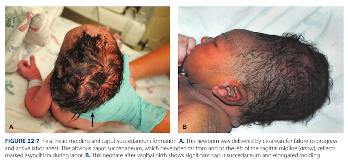
In occiput presentations, labor forces alter fetal head shape. In prolonged labors before complete cervical dilation, the portion of the fetal scalp immediately over the cervical os becomes edematous. This swelling is called caput succedaneum. It usually attains a thickness of only a few millimeters, but in prolonged labors it may be sufficiently extensive to prevent differentiation of the various sutures and fontanels. More commonly, the caput is formed when the head is in the lower portion of the birth canal and frequently only after the resistance of a rigid vaginal outlet is encountered. Because it develops over the most dependent area of the head, one may deduce the original fetal head position. In cases of marked asynclitism and dystocia, the caput succedaneum may form far from the sagittal midline (Fig. 22-7).
Molding refers to changes in the bony fetal head shape as a result of external compressive forces (see Fig. 22-7B). Possibly related to Braxton Hicks contractions, some molding develops before labor. Despite these shape changes, most studies indicate that the parietal bones seldom overlap. A “locking” mechanism at the coronal and lambdoidal sutures actually prevents such overlapping (Carlan, 1991).
Molding yields a shortened suboccipitobregmatic diameter. Of greatest importance in women with contracted pelves or asynclitism, the degree to which the head can mold may determine the difference between vaginal and cesarean birth. Some older literature implicated severe head molding as a cause for possible cerebral trauma. Because of the many associated factors, for example, prolonged labor with fetal sepsis and acidosis, it is impossible to link molding to any alleged fetal or neonatal neurological sequelae. Most molding resolves within the week following delivery, although persistent cases have been described (Graham, 2006). Differentiation of molding, caput succedaneum, and cephalohematoma is discussed in Chapter 33 (p. 608).
NORMAL LABOR CHARACTERISTICS
The greatest impediment (trở ngại) to understanding normal labor is recognizing its start. The strict definition describes uterine contractions that bring about demonstrable (rõ ràng) cervical effacement and dilation. Pinpointing (xác định chính xác) when labor actually begins, however, is challenging because determination is retrospective (hồi cứu). Several methods are used to mark its start. Despite these, a recent systematic review of labor definitions emphasizes the lack of consensus (đồng thuận) regarding definitions of labor onset (Hanley, 2016).
One method defines onset as the clock time when painful contractions become regular. However, uterine activity that causes discomfort but that does not represent true labor may develop at any time during pregnancy. False labor eventually wanes (biến mất, chấm dứt) spontaneously (tự nhiên), whereas the contractions of true labor typically progress to an effective, regular pattern (mô hình). A second method defines the onset of labor as beginning at the time of admission to a labor unit. In the United States, admission for labor is frequently based on the extent of cervical dilation accompanied by painful contractions. If a woman has intact (nguyên vẹn) membranes, a cervical dilation of 3 to 4 cm or greater and frequent painful contractions are reasonably reliable diagnostic criteria.
■ First Stage of Labor
For labor progression, Friedman (1954) described a characteristic sigmoid pattern by graphing cervical dilation against time.
With Friedman’s concept, the first stage of labor contains three functional labor divisions (ba giai đoạn chức năng), each with its own physiological characteristics (Fig. 22-8). First, during the preparatory division, although the cervix dilates little, its connective tissue components change considerably (Chap. 21, p. 406). Sedation and conduction analgesia are capable of arresting this labor division ->> bị ảnh hưởng bởi thuốc an thần, giảm đau???
The dilatational division, during which dilation proceeds at its most rapid rate, is unaffected by sedation. Last, the pelvic division commences (bắt đầu) with the deceleration (giảm) phase of cervical dilation. The cardinal movements of labor take place principally during this pelvic division. In actual practice, however, the onset of the pelvic division is seldom (hiếm khi) clearly identifiable.

Two phases of cervical dilation are defined. The latent phase corresponds to the preparatory division, and the active phase to the dilational division. Friedman further subdivided the active phase into the acceleration phase, the phase of maximum slope, and the deceleration phase (Fig. 22-9).
Latent Phase
The onset of latent labor, as defined by Friedman (1972), is the point at which the mother perceives regular contractions. For most women, this phase ends once cervical dilation of 4 cm is achieved. This threshold may be clinically useful, for it defines dilation limits beyond which active labor can be expected. More recently, the American College of Obstetricians and Gynecologists and Society for Maternal-Fetal Medicine (2019c) has redefined active labor to begin at 6 cm. A fuller discussion of these labor changes is found in Chapter 23 (p. 436)

A prolonged (kéo dài) latent phase was defined as one exceeding 20 hours in the nullipara and 14 hours in the multipara by Friedman and Sachtleben (1963). These times corresponded to the 95th percentiles. Factors that affected latent phase duration include excessive sedation or epidural analgesia (liều cao giảm đau ngoài màng cứng); unfavorable cervical condition (cổ tử cung không thuận tiện), that is, thick, uneffaced, or undilated; and false labor.
Of women in latent labor who had been administered heavy sedation, 85 percent eventually entered active labor. In another 10 percent, uterine contractions ceased (ngừng), which suggests false labor. In the remaining 5 percent, an abnormal latent phase persisted (tồn tại) and required oxytocin stimulation. Amniotomy (tia ối) was discouraged because of the 10-percent incidence of false labor. Friedman (1972) later reported that latent phase prolongation did not adversely influence fetal or maternal morbidity or mortality rates. However, prolongation of the latent phase appears associated with longer active labor duration and greater likelihood (khả năng) of obstetrical interventions (Ängeby, 2018; Chelmow, 1993; ilden, 2020).
Active Phase
The progress of labor in nulliparas has particular significance because these curves all reveal a rapid change in the slope of cervical dilation rates between 3 and 5 cm (see Fig. 22-9). Thus, cervical dilation of 3 to 6 cm or more, in the presence of uterine contractions, can be taken to reliably represent the threshold for active labor. Similarly, these curves provide useful guideposts for labor management. Turning again to Friedman (1955), the mean duration of active-phase labor in nulliparas was 4.9 hours. But, the standard deviation of 3.4 hours is large, hence, the active phase was reported to have a statistical maximum of 11.7 hours. Indeed, rates of cervical dilation ranged from a minimum of 1.2 up to 6.8 cm/ hr. Friedman (1972) also found that multiparas progress somewhat faster in active-phase labor, with a minimum normal rate of 1.5 cm/hr. His analysis of active-phase labor concomitantly describes rates of fetal descent and cervical dilation (see Fig. 22-8).
Descent begins in the later stage of active dilation, commencing at 7 to 8 cm in nulliparas and becoming most rapid after 8 cm. Others have reassessed the Friedman labor curves. Zhang and associates (2010) studied electronic labor records from 62,415 parturients with spontaneous labor at term and vaginal birth. For primiparas, the median time to progress rom 4 to 5 cm was 1.3 hours, from 5 to 6 cm 0.8 hours, and thereafter, additional centimeters were gained approximately each 0.5 hours. They found that normal labor may take >6 hours to progress from 4 to 5 cm and >3 hours to progress from 5 to 6 cm dilation. Rates for multiparas were similar to nulliparas from 4 to 6 cm, but then, labor accelerated much faster in multiparas. Data from this study form the foundation for new guidelines regarding cesarean delivery indications for labor arrest put forth in the American College of Obstetricians and Gynecologists’ Obstetric Care Consensus (2019c). These are described in Chapter 23 (p. 436).
■ Second Stage of Labor
Tis stage begins with complete cervical dilation and ends with fetal delivery. The median duration is approximately 50 minutes for nulliparas and about 20 minutes for multiparas, but it is highly variable (Kilpatrick, 1989). In a woman of higher parity with a previously dilated vagina and perineum, two or three expulsive efforts after full cervical dilation may suffice to complete delivery. Conversely, in a woman with a contracted pelvis, with a large fetus, or with impaired expulsive efforts from conduction analgesia or sedation, the second stage may become abnormally long. Nulliparas with high starting fetal station may experience a longer second stage (Ashwal, 2021). Greater maternal body mass index does not interfere with second-stage labor (Carlhäll, 2013; Robinson, 2011).
■ Labor Duration
The normal duration of labor may be clouded by the many clinical variables that affect the conduct of labor in modern obstetrical units. Kilpatrick and Laros (1989) reported that the mean length of first- and second-stage labors approximates 9 hours in nulliparas without regional analgesia and that the 95th percentile upper limit was 18.5 hours. Corresponding times for multiparas were a mean of 6 hours and a 95th percentile maximum of 13.5 hours. These authors defined labor onset as the time when a woman recalled regular, painful contractions every 3 to 5 minutes that led to cervical change.
Spontaneous labor was analyzed in nearly 25,000 women delivered at term at Parkland Hospital in the early 1990s. Almost 80 percent of women were admitted with a cervical dilation of ≤5 cm. Parity—nulliparous versus multiparous— and cervical dilation at admission were significant determinants of spontaneous labor length. The median time from admission to spontaneous delivery for all parturients was 3.5 hours, and 95 percent of all women delivered within 10.1 hours. These results suggest that normal human active labor is relatively short.
MANAGEMENT OF NORMAL LABOR
Birthing is a normal physiological process that most women experience without complications. However, unexpected intrapartum (trong khi sinh) complications can arise quickly. Thus, every woman and her supporters should feel welcomed and comfortable, yet safety measures must be in place for the mother and her newborn if complications suddenly develop. The American Academy of Pediatrics and the American College of Obstetricians and Gynecologists (2017) have collaborated (hợp tác) in the development of Guidelines for Perinatal Care. These provide detailed inhormation on the appropriate content of intrapartum care, including both personnel (nhân sự) and facility (cơ sở vật chất) requirements. Labor and delivery outside the hospital is elected by some parturients (sản phụ). Tis option is discussed in Chapter 27 (p. 504).
■ Emergency Medical Treatment and Labor
Act—EMTALA
By law, all Medicare-participating hospitals with emergency services must provide an appropriate screening examination for any pregnant woman experiencing contractions and presenting to the emergency department for evaluation. The definition of an emergency condition makes specific reference to a pregnant woman who is having contractions. Labor is defined as “the process of childbirth beginning with the latent phase of labor continuing through delivery of the placenta. A woman experiencing contractions is in true labor unless a physician certifies that after a reasonable time of observation the woman is in false labor.” A woman in true labor is considered “unstable” for interhospital transfer purposes until the newborn and placenta are delivered. A stable woman may, however, be transferred at the direction of the patient or by a physician who certifies that the benefits of treatment at another facility outweigh the transfer risks.
■ Identification of Labor
Pregnant women are urged to report early in labor rather than to procrastinate (trì hoãn) until delivery is imminent (sắp xảy ra). Early admittance is important if during antepartum (trước sinh) care, the woman, her fetus, or both are found to carry risks for intrapartum complications.
Although the differentiation between false and true labor is difficult at times, the diagnosis usually can be clarified (làm rõ) by cervical dilation and by contraction frequency and intensity. Pates and associates (2007) studied one commonly used recommendation given to pregnant women. Namely (cụ thể), in the absence of ruptured membranes or bleeding, uterine contractions 5 minutes apart for 1 hour—that is, ≥12 contractions in 1 hour— may signify labor onset. Among 768 women in this study at Parkland Hospital, active labor defined as cervical dilation ≥4 cm was diagnosed within 24 hours in three fourths of women with ≥12 contractions per hour.
In those instances (ví dụ) when a diagnosis of labor cannot be established with certainty, observation for a longer period is reasonable. Women who present to Parkland Hospital for labor symptoms at ≥240/7 weeks gestation are routinely evaluated in a labor triage unit (đơn vị phân loại chuyển dạ), which is contiguous (tiếp giáp) to our labor and delivery unit. All women in the triage unit are evaluated by nurse practitioners and certified nurse midwives using written protocols. For those with uncomplicated pregnancies, women with intact membranes and cervical dilation <4 cm receive continuous external fetal monitoring for up to 2 hours. Women diagnosed with labor by either cervical change or persistent (kéo dài) uterine contractions are admitted. After review by a physician, women without cervical change or with abatement (giảm) of contractions return home with a diagnosis of false labor. In a recent study, a total of 3949 women with uncomplicated pregnancies between 370/7 and 416/7 weeks’ gestation were diagnosed with false labor. The mean interval (khoảng thời gian trung bình) from hospital discharge to when they again presented was 4.9 days (Nelson, 2017). Within this protocol, hospital discharge with false labor at term was not associated with higher rates of adverse neonatal outcomes or cesarean delivery. The American College of Obstetricians and Gynecologists (2020a) has endorsed hospital-based obstetrical triage units.
Instead of false labor, labor in the latent phase (pha tiềm thời) may be diagnosed. In this phase, some women may require admission for pain management. However, for women without antepartum risks and with reassuring maternofetal status, expectant care during latent labor is reasonable (American College of Obstetricians and Gynecologists, 2019a). Studies support the value of this approach to potentially lower rates of interventions that include oxytocin, antibiotics for intrapartum fever, and cesarean delivery (Bailiet, 2005; Wood, 2016). Instructions (hướng dẫn) on coping (ứng phó) and self-care during this time have value (Hosek, 2014). Moreover, a mutually agreed upon time is set for reevaluation.
■ Initial Evaluation
For the gravida (thai phụ) presenting with symptoms of labor, blood pressure, temperature, pulse, and respiratory rate are recorded. Fetal heart rate is evaluated using a portable (cầm tay) Doppler device, sonography, or fetoscope. Our practice uses extended electronic fetal monitoring to assess initial fetal status. The pregnancy record is promptly reviewed to identify complications. A vaginal examination is performed unless there is a known placenta previa or vasa previa. The gloved index and second fingers are introduced into the vagina while avoiding the anal region.
Ruptured Membranes
During prenatal care, the woman is instructed to be aware of fluid leakage from the vagina and to report this event promptly. Rupture of the membranes is significant for three reasons. First, if the presenting part is not flexed in the pelvis, the umbilical cord can prolapse and be compressed. Second, labor is likely to begin soon if the pregnancy is at or near term. Third, if delivery is delayed after membrane rupture, intrauterine and neonatal infection is more likely as the time interval lengthens (Herbst, 2007).
During sterile speculum examination, ruptured membranes are diagnosed if amnionic fluid pools in the posterior fornix or clear fluid flows from the cervical canal. If the diagnosis remains uncertain, one method involves pH determination of vaginal fluid. The pH of vaginal secretions normally ranges from 4.5 to 5.5, whereas the pH of amnionic fluid is usually >7.0. The use of the indicator nitrazine to identify ruptured membranes is a simple and fairly reliable method. Test papers are impregnated with the dye, and the color of the reaction between these paper strips and vaginal fluids is interpreted by comparison with a standard color chart posted on the strip’s container. A pH above 6.5 is consistent with ruptured membranes. False-positive test results may stem from blood, semen, or bacterial vaginosis, which raise pH. Scant fluid can yield a false-negative test result.
Another test to identify amnionic fluid microscopically evaluates vaginal fluid that is dried on a slide. Arborization or a tern pattern suggests amnionic rather than cervical fluid. Amnionic fluid crystallizes and erns due to its concentrations of sodium chloride, proteins, and carbohydrates. Of other methods, detection of alpha-fetoprotein in the vaginal vault has been used to identify amnionic fluid (Yamada, 1998). Rarely required, identification may also follow injection of 1 to 4 mL of a 5-percent sodium fluorescein solution into the amnionic sac via abdominal amniocentesis (Ireland, 2017).
Last, specific amnionic fluid proteins can be sought using point-of-care assays. These include AmniSure, which binds placental alpha microglobulin-1; Actim PROM, with binds insulin growth factor binding protein-1 (IGFBP-1); and ROM Plus, which detects IGFBP-1 plus alpha-fetoprotein (Eleje, 2017; Palacio, 2014). False-positive and false-negative results are not uncommon. Te Food and Drug Administration (2018) emphasizes the need to incorporate clinical findings if these methods are used.
Cervical Assessment
Progress during labor produces specific cervical changes. Cervical dilation is assessed by sweeping the examining finger from the inner cervical rim on one side of the opening to the opposite inner rim. The diameter traversed is estimated in centimeters. Effective labor will dilate the cervix, which is considered fully dilated when the diameter measures 10 cm. The presenting part of a term-size newborn usually can pass through a cervix this widely dilated.
Cervical effacement is the length of the cervical canal compared with that of an unlabored cervix. When the length of the cervix is reduced by one half, it is 50-percent efaced. When the cervix becomes as thin as the adjacent lower uterine segment, it is completely, or 100-percent, efaced. Cervical position reflects the relationship of the cervical os to the center point of the presenting part. It is categorized as posterior, midposition, or anterior. Fetal descent will gradually move the cervix anteriorly. Along with position, the consistency of the cervix is determined to be soft, firm, or intermediate between these two. Cervical softening aids dilation and effacement.
Fetal station describes the presenting fetal part’s leading edge in the birth canal in relationship to the ischial spines. These spines lie halfway between the pelvic inlet and the pelvic outlet and lie at the level of the midpelvis. When the lowermost portion of the presenting fetal part reaches the spines, it is designated as being at zero (0) station.
In the past, the long axis of the birth canal above and below the ischial spines was arbitrarily divided into thirds by some and into ths (approximately 1 cm) by other groups. In 1989, the American College of Obstetricians and Gynecologists adopted the classification of station that divides the pelvis above and below the spines into ths. Each th represents 1 cm above or below the spines. Thus, as the presenting fetal part descends from the inlet toward the ischial spines, the designation is –5, –4, –3, –2, –1, and then 0 station. Below the spines, as the presenting fetal part descends, it passes +1, +2, +3, +4, and +5 stations to delivery. Station +5 cm corresponds to the fetal head being visible at the introitus.
If the leading part of the fetal head is at 0 station or below, most often the fetal head has engaged—thus, the biparietal plane has passed through the pelvic inlet. If the head is unusually molded or if caput succedaneum formation is extensive, or both, engagement might not have taken place although the scalp is at 0 station.
In addition to charting the progress of labor, these five characteristics—cervical dilation, effacement, consistency, position, and fetal station—are assessed when tabulating the Bishop score. This score is commonly used to predict labor induction outcome and is discussed in Chapter 26 (p. 488). Taken together, these factors suggest the subjective “favorability” of the cervix for induction success.
Laboratory Studies
When a woman is admitted in labor, most often the hematocrit or hemoglobin concentration is rechecked. The hematocrit can be measured easily and quickly. At Parkland Hospital, blood is collected in a standard collection tube with anticoagulant.
From this, a heparinized capillary tube is filled to spin in a microhematocrit centrifuge in the labor and delivery unit. This provides a hematocrit value within 3 minutes. The initial collection tube is also sent to the hematology laboratory for evaluation if the point-of-care hematocrit is <30 volume percent. Another labeled tube of blood is allowed to clot and sent to the blood bank for blood type and antibody screen. Some states, for example, Texas, require routine testing for syphilis, hepatitis B, and HIV in all women admitted to labor and delivery units, even if these were done during prenatal care. In some labor units, a clean-catch voided specimen is examined in all women for protein and glucose. At Parkland Hospital, we obtain a urine specimen for protein determination in hypertensive women only (Chap. 40, p. 689).
■ Management of First-stage Labor
As soon as possible after admittance, the remainder of a general examination is completed. Whether a pregnancy is normal can best be determined when all examinations, including record and laboratory review, are completed. A rational plan for monitoring labor can then be established based on the needs of the fetus and the mother. Because labor lengths vary markedly among individuals, precise statements to the patient regarding anticipated labor duration are unwise.
Intrapartum Fetal Monitoring
This is discussed in detail in Chapter 24. Briefly, the American Academy of Pediatrics and American College of Obstetricians and Gynecologists (2017) recommend that during first-stage labor, in the absence of any abnormalities, the fetal heart rate should be checked immediately after a contraction at least every 30 minutes and then every 15 minutes during second-stage labor. If continuous electronic monitoring is used, the tracing is evaluated at least every 30 minutes during the first stage and at least every 15 minutes during the second stage. For women with pregnancies at risk, fetal heart auscultation is performed at least every 15 minutes during first-stage labor and every 5 minutes during the second stage. Continuous electronic monitoring may be used with evaluation of the tracing every 15 minutes during the first stage of labor, and every 5 minutes during the second stage.
Maternal Monitoring
Temperature, pulse, and blood pressure are evaluated at least every 4 hours. If membranes have been ruptured for many hours or if there is a borderline temperature elevation, the temperature is checked hourly. Although uterine contractions are usually assessed with electronic monitoring, they can be quantitatively and qualitatively evaluated manually. With the palm of the hand resting lightly on the uterus, the time of contraction onset is determined. Its intensity is gauged from the degree of muscle tone the uterus achieves. At the acme of effective contractions, the finger or thumb cannot readily indent the uterus during a “firm” contraction. The time at which the contraction disappears is noted next. This sequence is repeated to evaluate the frequency, duration, and intensity of contractions.
During the first stage of labor, the need for subsequent vaginal examinations to monitor cervical change and presenting part position will vary considerably. When the membranes rupture, an examination to exclude cord prolapse is performed expeditiously if the fetal head was not definitely engaged at the previous examination. The fetal heart rate is also checked immediately and during the next uterine contraction to help detect occult umbilical cord compression. At Parkland Hospital, periodic pelvic examinations are typically performed at 2- to 3-hour intervals for gravidas in active labor to evaluate labor progress. Evidence implicating the number of vaginal examinations in infection-related morbidity is conflicting (Cahill, 2012; Soper, 1989).
Maternal Position
In bed, the laboring woman may assume the position she finds most comfortable, and often this will be lateral recumbency. Lying supine is typically avoided to avert aortocaval compression and its potential to lower uterine perfusion (Chap. 4, p. 64). However, the normal laboring woman need not be confined to bed early in labor. A comfortable chair may be beneficial psychologically and perhaps physiologically. Others encourage ambulation.
Proponents of walking report that it shortens labor, lowers rates of oxytocin augmentation, diminishes the need for analgesia, and decreases the frequency of operative vaginal delivery (Flynn, 1978; Read, 1981). In their Cochrane review, Lawrence and associates (2013) reported that labor in ambulant or upright positions shortened first-stage labor by approximately 1 hour and lowered cesarean delivery and epidural analgesia rates. However, they cautioned interpretation given variable study quality. Lupe and Gross (1986) concluded, however, that no conclusive evidence supports assertions that upright maternal posture or ambulation improves labor. They reported that women preferred to lie on their side or sit in bed. Few chose to walk, fewer to squat, and none wanted the knee-chest position.
Parturients tended to assume fetal positions in later labor. Most women enthusiastic about ambulation returned to bed when active labor began (Carlson, 1986; Williams, 1980). Bloom and colleagues (1998) conducted a randomized trial to study the effects of walking during first-stage labor. In 1067 women with uncomplicated term pregnancies delivered at Parkland Hospital, these investigators reported that ambulation did not affect labor duration. Ambulation did not reduce the need for analgesia nor was it harmful to the newborn. Because of these observations, we give women without complications the option to select either recumbency or supervised ambulation during labor.
Pain Management
In general, the needs and desires for pain relief are directed by the parturient. Relaxation techniques may have a role for pain relief and patient satisfaction, although evidence-based data are not robust (Smith, 2018). Discussed in the last section, ambulation is another option. Some women choose to spend part of first-stage labor in a large water tub, if available, for pain relief.
With this practice, one Cochrane review found lower rates of regional analgesia use and no greater adverse neonatal or maternal effects compared with traditional labor (Cluett, 2018). Pharmacologic options include epidural analgesia; intermittent intravenous (IV) or intramuscular (IM) opioids or meperidine (Table 25-2, p. 469), and less often, nitrous oxide. These are discussed in detail in Chapter 25.
Oral Intake
Food and liquids with particulate matter should be withheld during active labor and delivery. Gastric emptying time is remarkably prolonged once labor is established and analgesics are administered. As a consequence, ingested food and most medications remain in the stomach and are poorly absorbed. They may be vomited and aspirated (Chap. 25, p. 482). However, oral intake of moderate amounts of clear liquids is reasonable for women with uncomplicated labor (American Academy of Pediatrics, 2017; American Society of Anesthesiologists, 2016). Water, clear tea, black coffee, carbonated beverages, Popsicles, and pulp-free juices are options. In those with appreciable risks for aspiration or those with significant risks for cesarean delivery, further restriction may be instituted. For example, for those with planned cesarean delivery, liquids are halted 2 hours before and solids are stopped 6 to 8 hours prior to surgery (American College of Obstetricians and Gynecologists, 2019b).
Intravenous Fluids
Although an IV infusion system is often routinely established early in labor, real need for this in the normal pregnant woman is limited. Access allows parenteral analgesia or IV fluid infusion prior to regional analgesia. In the immediate puerperium, a dilute oxytocin solution can be given prophylactically to prevent uterine atony and at times therapeutically to treat it.
Moreover, with longer labors, the administration of glucose, sodium, and water to the otherwise fasting woman at the rate of 60 to 120 mL/hr prevents dehydration and acidosis. Although not mandatory, IV hydration may shorten labor length, and most studies of fluid selection use first-stage labor length as a primary outcome. From these investigations, data support use of fluids containing dextrose compared with fluid without it (Riegel, 2018; Shrivastava, 2009). For administration, rates of 125 or 250 mL/hr are suitable (Dawood, 2013; Ehsanipoor, 2017; Fong, 2017). In our practice, saline with 5-percent dextrose is infused at a rate o 150 mL/hr.
Rupture of Membranes
If the membranes are intact, temptation is great, even during normal labor, to perform amniotomy. Benefits are earlier detection of meconium-stained amnionic fluid and the opportunity to apply an electrode to the fetus or insert a pressure catheter into the uterine cavity for monitoring. In one metaanalysis of 15 studies, normal spontaneous labor was not shortened by amniotomy (Smyth, 2013). Risks include potential uterine infection if performed early and possible cord prolapse. To help prevent prolapse, the fetal head must be well applied to the cervix and not dislodged from the pelvis during amniotomy. The use of amniotomy in the setting of abnormal labor is discussed in Chapter 23 (p. 434).
With prolonged membrane rupture and unknown group B streptococcal (GBS) status, antimicrobial administration for prevention of GBS infections is recommended for intrapartum risk factors (American College of Obstetricians and Gynecologists, 2020b). These include rupture of membranes greater than 18 hours or intrapartum temperature >38.0°C or >100.4°F (Chap. 67, p. 1194). Tis practice similarly lowers rates of chorioamnionitis and endometritis (Saccone, 2015).

Urinary Bladder Function
Distention of the bladder can hinder descent of the fetal presenting part and lead to subsequent bladder hypotonia and infection. Periodically, the suprapubic region is palpated to detect distention. If the bladder is readily seen or palpated above the symphysis, the woman should be encouraged to void. Tose unable to do so on a bedpan may be able to ambulate with assistance to a toilet. Catheterization is indicated if the bladder is distended and voiding is not possible. Regional analgesia is a common reason. In these cases, continuous or intermittent catheterization is suitable, and both have comparable rates of puerperal urinary tract infection and urinary retention (Li, 2019).
Postpartum retention is discussed in Chapter 36 (p. 643).
■ Management of Secondstage Labor

In later first-stage or early second-stage labor, attempts are made to determine head position. The fingers are directed posteriorly and then swept forward over the fetal head toward the maternal symphysis (Fig. 22-10). During this movement, the fingers necessarily cross the sagittal suture, and its lie is delineated. Next, the positions of the two fontanels are ascertained.
For this, fingers are passed to the most anterior extension of the sagittal suture, and the fontanel encountered there is examined and identified. The fingers then pass along the sagittal suture to the other end of the head until the other fontanel is felt and differentiated (Fig. 22-11). Last, the station, or extent to which the presenting part has descended into the pelvis, also can be established. Using these maneuvers, the various sutures and fontanels are determined. In unclear cases, sonography can be used as described earlier (p. 420).
With full cervical dilation, which signifies the onset of the second stage, a woman typically begins to bear down. With descent of the presenting part, she develops the urge to defecate. Uterine contractions and the accompanying expulsive forces may now last 1 minute and recur at intervals less than 90 seconds. As discussed earlier, the median duration of the second stage is 50 minutes in nulliparas and 20 minutes in multiparas, although this length can vary. Monitoring intervals of the fetal heart rate were discussed earlier (p. 427), and interpretation of second-stage electronic fetal heart rate patterns is discussed in Chapter 24 (p. 447).
In most cases, bearing down is reflexive and spontaneous during second-stage labor. In those with neuraxial analgesia, this urge may be blunted. To improve spontaneous birth rates, delayed pushing for 60 minutes once the second stage is reached has been suggested to permit passive fetal descent to increase pushing efficiency and minimize maternal exhaustion. One large randomized trial compared immediate and delayed pushing in nulliparas at term with neuraxial analgesia. Routes of delivery were similar. The immediate group actively pushed longer, a mean difference of 9 minutes, but had lower rates of chorioamnionitis (Cahill, 2018). A subsequent metaanalysis noted similar findings (Di Mascio, 2020).
During pushing, a woman may not employ her expulsive forces to good advantage and coaching is desirable.
When the next uterine contraction begins, she is instructed to exert downward pressure as though she were straining at stool. A woman is not encouraged to push beyond the completion of each contraction. Instead, she and her fetus are allowed to rest and recover. During this period of active pushing, the fetal heart rate auscultated during the contraction is likely to be slow but should recover to normal range beore the next expulsive effort. Fetal and obstetrical outcomes appear to be unaffected whether pushing is coached or uncoached during second-stage labor (Bloom, 2006; uuli, 2012).
Several positions during the second stage have been recommended to augment pushing efforts. The Birth in the Upright Maternal Position with Epidural in Second Stage (BUMPES) trial compared a lateral recumbent position and a supported upright one for nulliparas. Upright positions include standing, sitting out of bed, kneeling, or squatting. The lateral recumbent position required the head of the bed to have an incline ≤30 degrees. When compared, the recumbent group had a spontaneous birth rate o 41 percent, which was significantly higher than the 35-percent spontaneous birth rate experienced by the upright group. Otherwise, rates of operative vaginal delivery (OVD), anal sphincter tear, and neonatal outcome measures were similar (Epidural and Position trial Collaborative Group, 2017).
In a subsequent Cochrane review of studies that included nulliparas and multiparas, upright or recumbent position had little or no affect on rates of “operative birth,” which combined OVD and cesarean delivery (Walker, 2018). In those without epidural analgesia, Gupta and coworkers (2017) in their Cochrane review compared upright positions with supine or lithotomy positions during second-stage labor in multiparas and nulliparas. Upright positions offered a marginally shorter interval to delivery and fewer episiotomies and OVDs. However, rates of blood loss >500 mL and perhaps of second-degree lacerations were increased. In sum, no one position is mandated for labor. Repositioning with the goal of maternal comfort and optimal fetal trajectory is suitable as long as appropriate maternal and fetal monitoring can be achieved (American College of Obstetricians and Gynecologists, 2019a). To help avoid femoral or lumbosacral nerve injury from lithotomy positions, hips are not overly flexed, abducted, or externally rotated for extended periods (Chap. 36, p. 644). From early data, transperineal ultrasound assessment of the fetal head’s angle of progression relative to the symphysis during the second stage may be a tool to predict vaginal birth (Nassr, 2021).
As the head descends through the pelvis, the perineum begins to bulge and the overlying skin becomes stretched. Now the scalp of the fetus may be visible through the vulvar opening. At this time, the woman and her fetus are prepared for delivery, which is described in Chapter 27 (p. 498).
LABOR MANAGEMENT PROTOCOLS
An orderly and systematic approach to labor management results in reproducible beneficial maternal and perinatal outcomes (Althabe, 2008). In Dublin more than 30 years ago, O’Driscoll and associates (1984) pioneered the concept that a disciplined, standardized labor management protocol reduced the number of cesarean deliveries for dystocia. Their overall cesarean delivery rate was 5 percent in the 1970s and 1980s with such management. The approach is now referred to as active management of labor. Two of its components—amniotomy and oxytocin— have been widely used, especially in English-speaking countries outside the United States. With this protocol, labor is diagnosed when painful contractions are accompanied by complete cervical effacement, bloody “show,” or ruptured membranes.
Women with such findings are committed to delivery within 12 hours. Pelvic examination is performed each hour for the next 3 hours, and thereafter at 2-hour intervals. When dilation has not increased by at least 1 cm/hr, amniotomy is performed. Progress is again assessed at 2 hours, and high-dose oxytocin infusion, described in Chapter 26 (p. 493), is started unless dilation of at least 1 cm/hr is attained. Women are constantly attended by midwives. If membranes rupture before admission, oxytocin is begun for no progress at the 1-hour mark. López-Zeno and colleagues (1992) prospectively compared such active management with their “traditional” approach to labor management at Northwestern Memorial Hospital in Chicago. They randomly assigned 705 nulliparas with uncomplicated pregnancies in spontaneous labor at term. The cesarean delivery rate was significantly lower with active versus traditional management—10.5 versus 14.1 percent, respectively. Subsequent studies did not show this. Wei and associates (2013) in a Cochrane database review found a modest reduction in cesarean delivery rates when active management of labor was compared with standard care. Frigoletto and coworkers (1995) reported another randomized trial with 1934 nulliparous women at Brigham and Women’s Hospital in Boston. Although they found that such management somewhat shortened labor, it did not affect the cesarean delivery rate. These observations have since been reported by others (Brown, 2008).
At Parkland Hospital, women are admitted if active labor is diagnosed or if ruptured membranes are confirmed. Labor is defined as cervical dilation of ≥4 cm in the presence of regular uterine contractions. Management guidelines direct that a pelvic examination subsequently be performed approximately every 2 hours. Ineffective labor is suspected when the cervix does not dilate within approximately 2 hours of admission. Amniotomy is then performed, and labor progress determined at the next 2-hour evaluation. In women whose labors do not progress, an intrauterine pressure catheter is placed to assess uterine function. Hypotonic contractions and no cervical dilation after an additional 2 to 3 hours result in stimulation of labor using the high-dose oxytocin regimen described in Chapter 26 (p. 493).
The goal is uterine activity of 200 to 250 Montevideo units for 4 hours before dystocia is diagnosed. In cases in which hypotonic contractions are strongly suspected, internal monitors may be placed with amniotomy and again cervical change and contraction pattern are assessed in 2 hours. Confirmation of deficient Montevideo units at that time may prompt oxytocin augmentation for maternal or fetal indications.
Dilation rates of 1 to 2 cm/hr are accepted as evidence of progress after satisfactory uterine activity has been established with oxytocin. This can require up to 8 hours or more before cesarean delivery is performed for dystocia. The cumulative time required to efect this stepwise management approach permits many women to establish effective labor. This management protocol has been evaluated in >20,000 women with uncomplicated pregnancies. Importantly, these labor interventions and the relatively infrequent use of cesarean delivery did not jeopardize the fetus-newborn.
Nhận xét
Đăng nhận xét